Sub-Ångstrom 3D Resolution,
Large-Volume Imaging
and Automation Advances
in Electron Ptychography


Outline

Motivation: “We can compute what we cannot yet see”
The gap between atomistic simulations and 3D atomic resolution imaging
- ML potentials are now probing 108 atoms at picosecond–nanosecond time‑steps
- 2024: [1] polycrystalline Cu, 100M atoms
- 2023: [2] SARS-CoV-2 Spike glycoprotein in water, 100M atoms
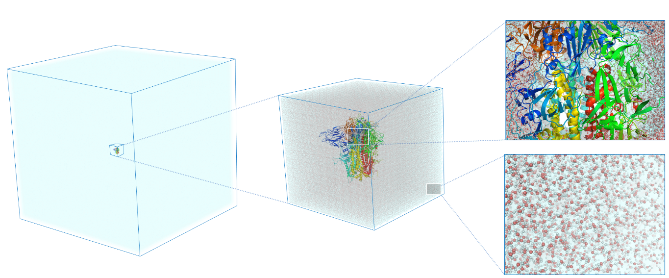
- 3D atomic resolution imaging has been limited to max. volumes of ~(15nm)3, 104 - 105 atoms
![]()
![]()
Adapted from [3]

Note
Ptychographic electron tomography can be developed into an experimental counterpart that can shorten this gap by combining ML-enhanced reconstructions, automation & dose‑efficient scanning.

Successive approximations in STEM ptychography

\(\psi_{out} = (1+i\phi)\psi_{in}\)
\(\psi_{out} = A(\vec{r}) \cdot e^{i\sigma V(\vec{r})} \cdot \psi_{in}\)
\(\psi_{out} = \mathcal{M}_V(\psi_{in})\)
✅ Real-time results & calibration
✅ Fast reconstruction for thin samples
✅ 2.5D reconstruction for thick samples
✅ highest x-y resolution
Note
Each approximations has its use in ptychographic tomography
Our protagonists today: nanoscale particles and quantum materials
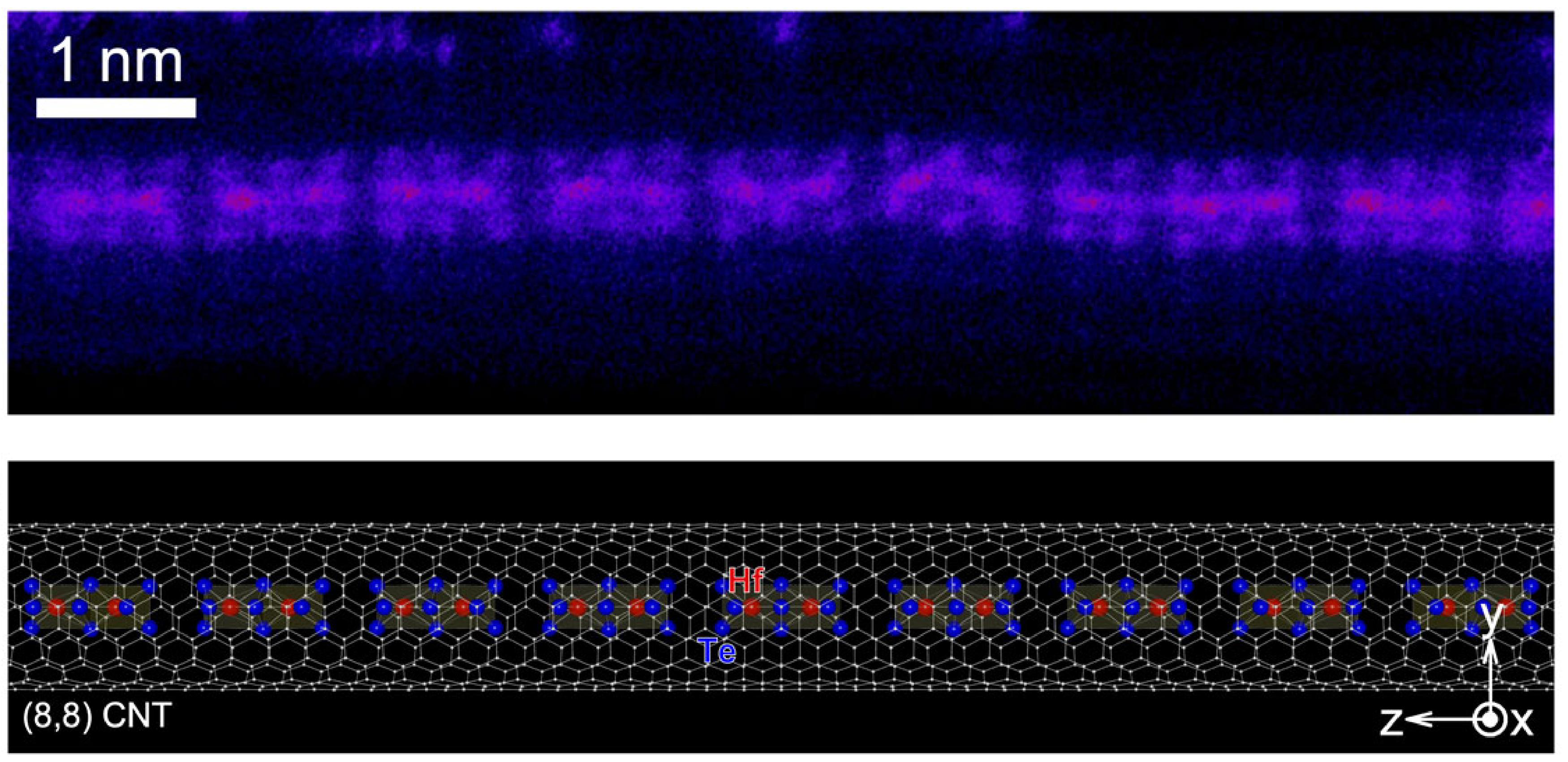
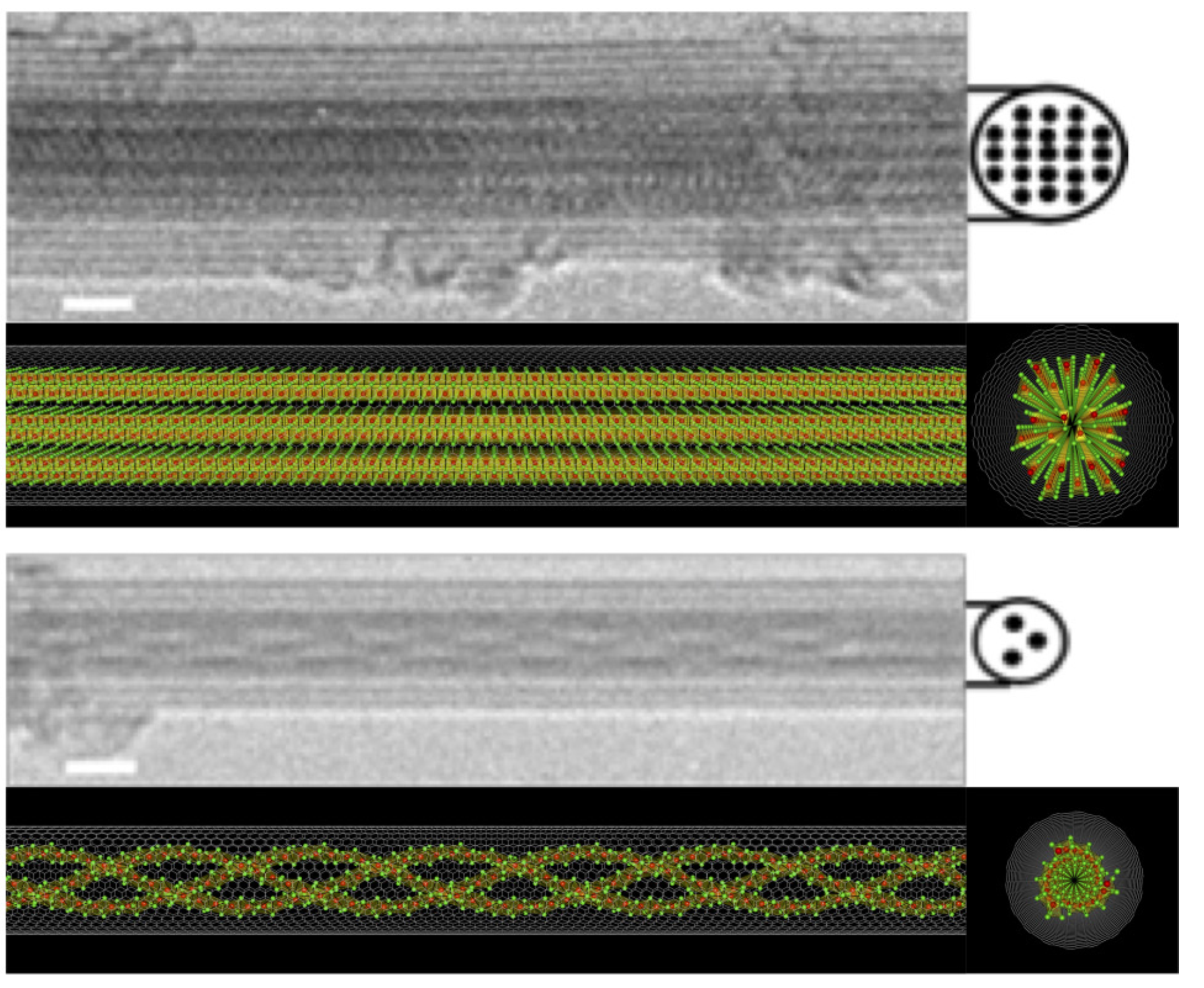
1️⃣ Transition metal chalcogenides in the few-chain limit
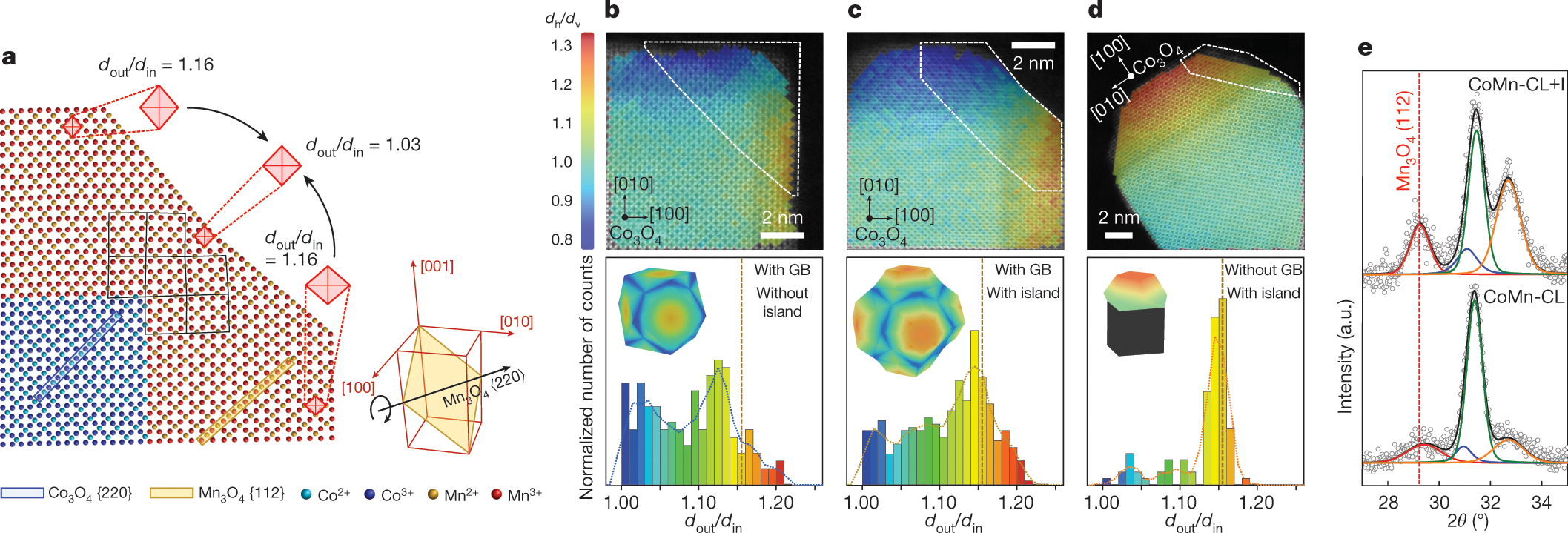
2️⃣ Strain-engineered Core-Shell Oxide particles
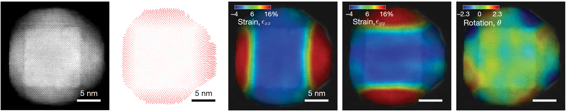
3️⃣ Strain-engineered chiral nanoparticles
Note
Quantum materials and nanoparticles are ideal test cases for our atomic resolution 3D imaging as we develop scalable algorithms
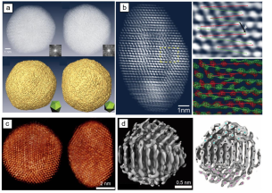
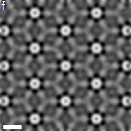
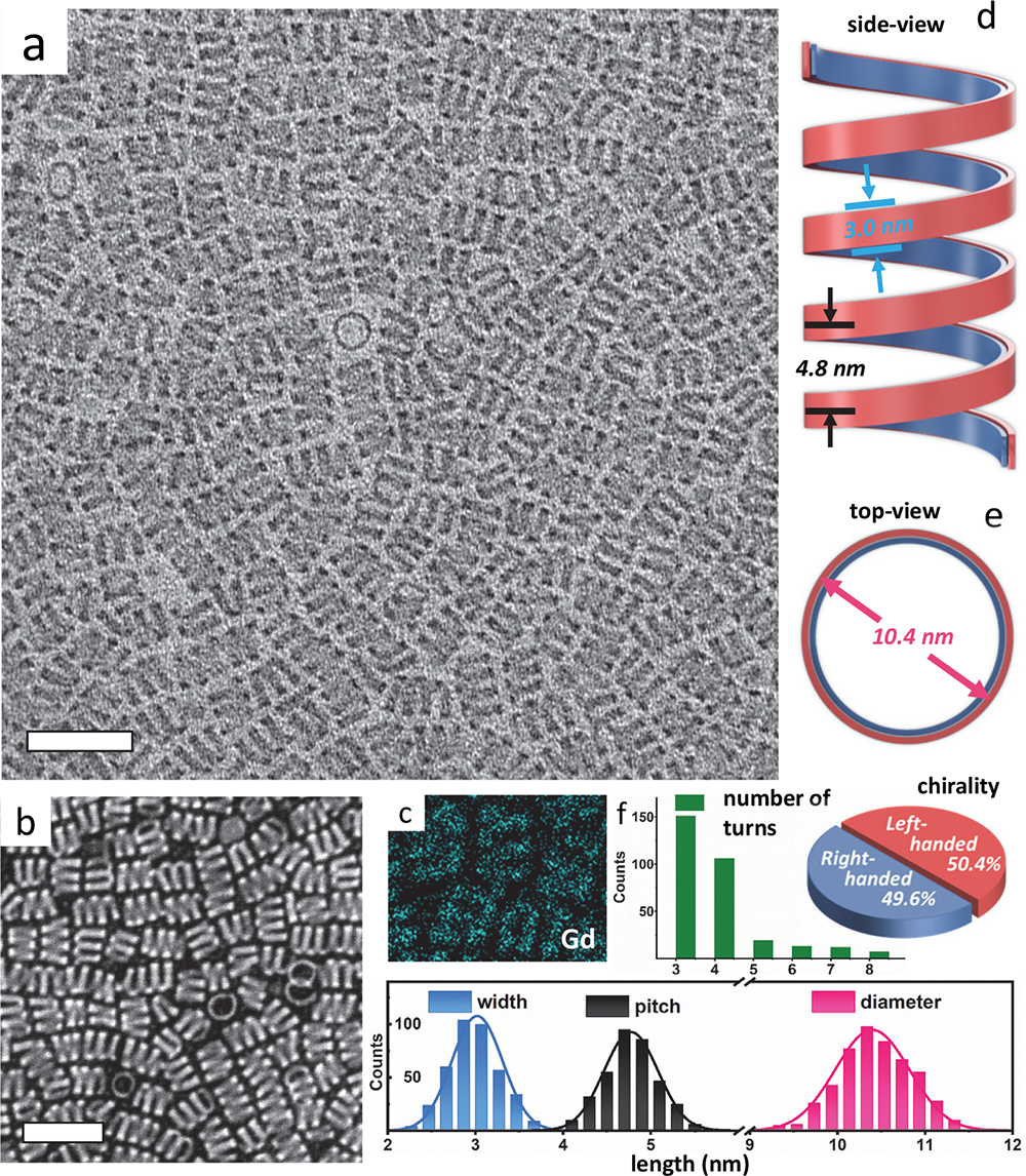
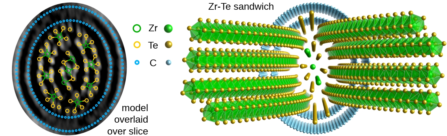
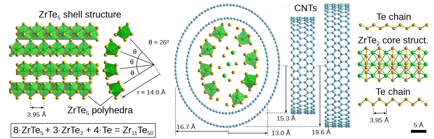
Ptychographic Tomography Solves Nanostructures
Note
First 3D atomic structure solved with phase-contrast tomography.
Novel ZrTe2 phase, confirmed stable with DFT simulations.
Depth Resolution Progress Over Time
Note
Algorithm development drives resolution records and depth of focus enhancements
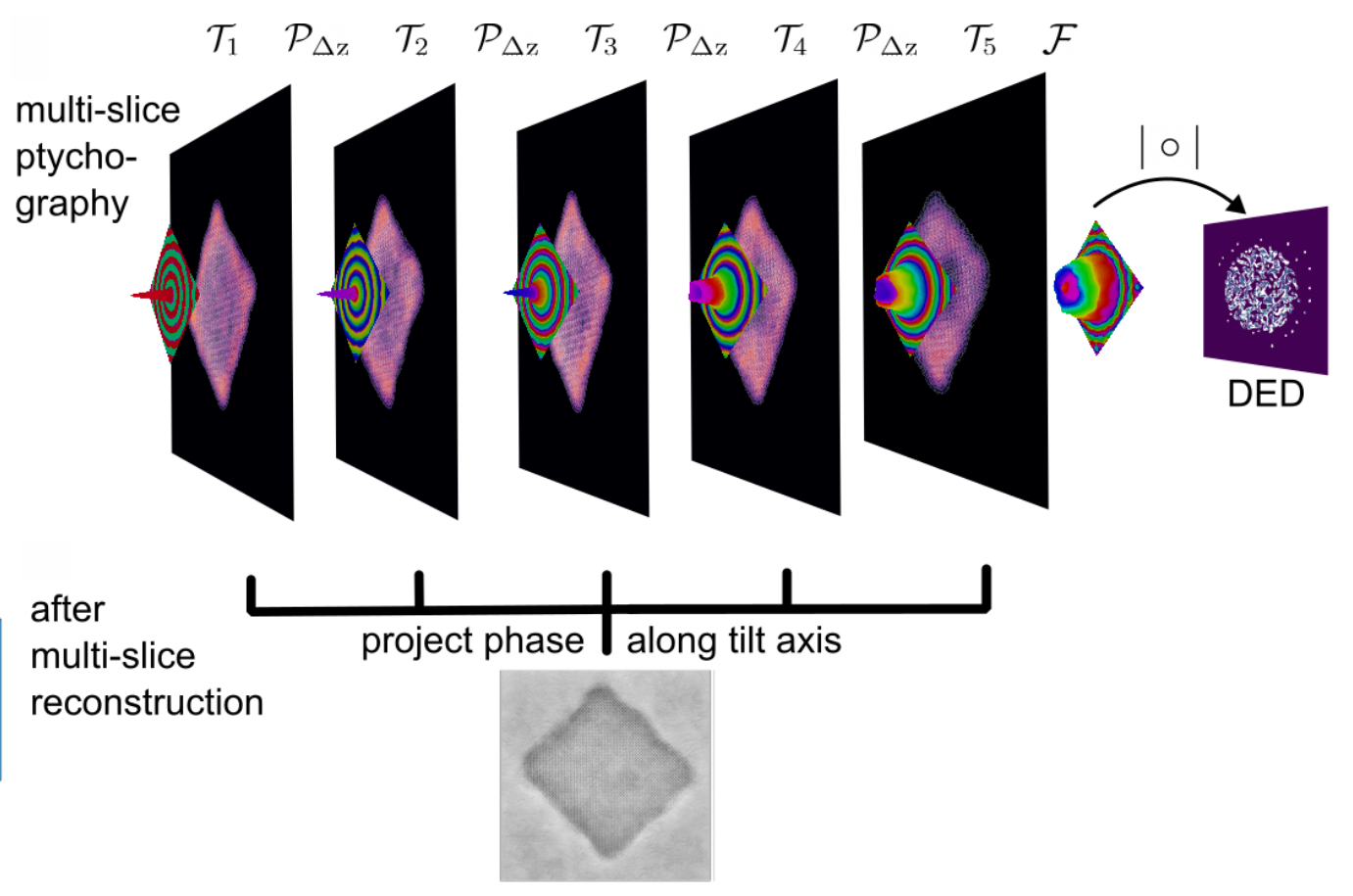
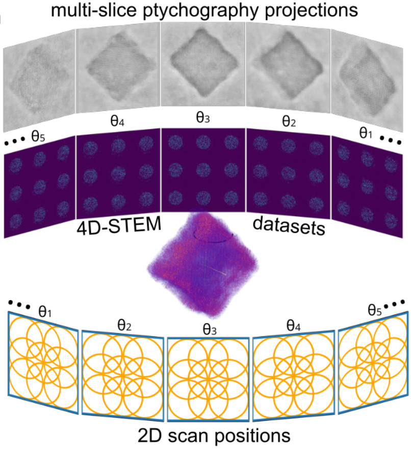
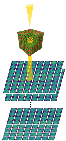
Next step: Multi-Slice Ptychographic Tomography
for each tilt angle
and project the potential along z
✅ Advantages:
- Decouple tomographic alignment from ptychographic reconstruction
- Can use positions and alignment as input to E2E-MSPT
❌ Disadvantage:
- Loses some 3D info from MSP

Joint Tomography and Rigid Alignment enables atomic resolution of beyond-DOF volumes
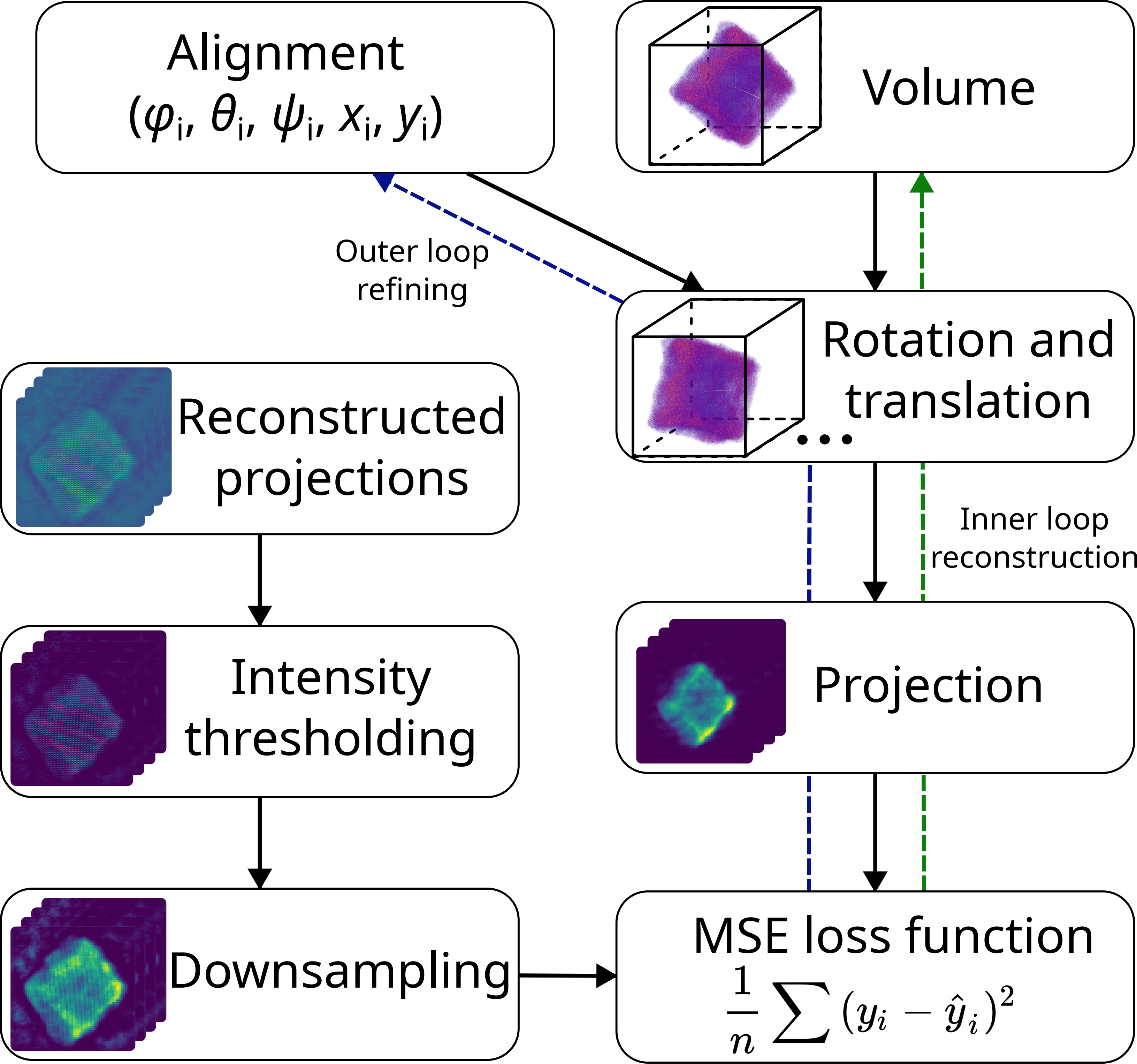
Outer loop: rigid alignment
Inner loop: fixed-alignment reconstruction
Note
Enabled by reaching sub-pixel alignment at each scale
3x DOF volumes display atomic resolution
Note
Volume size: (18.2 nm)3 Voxel size: 0.3 Å
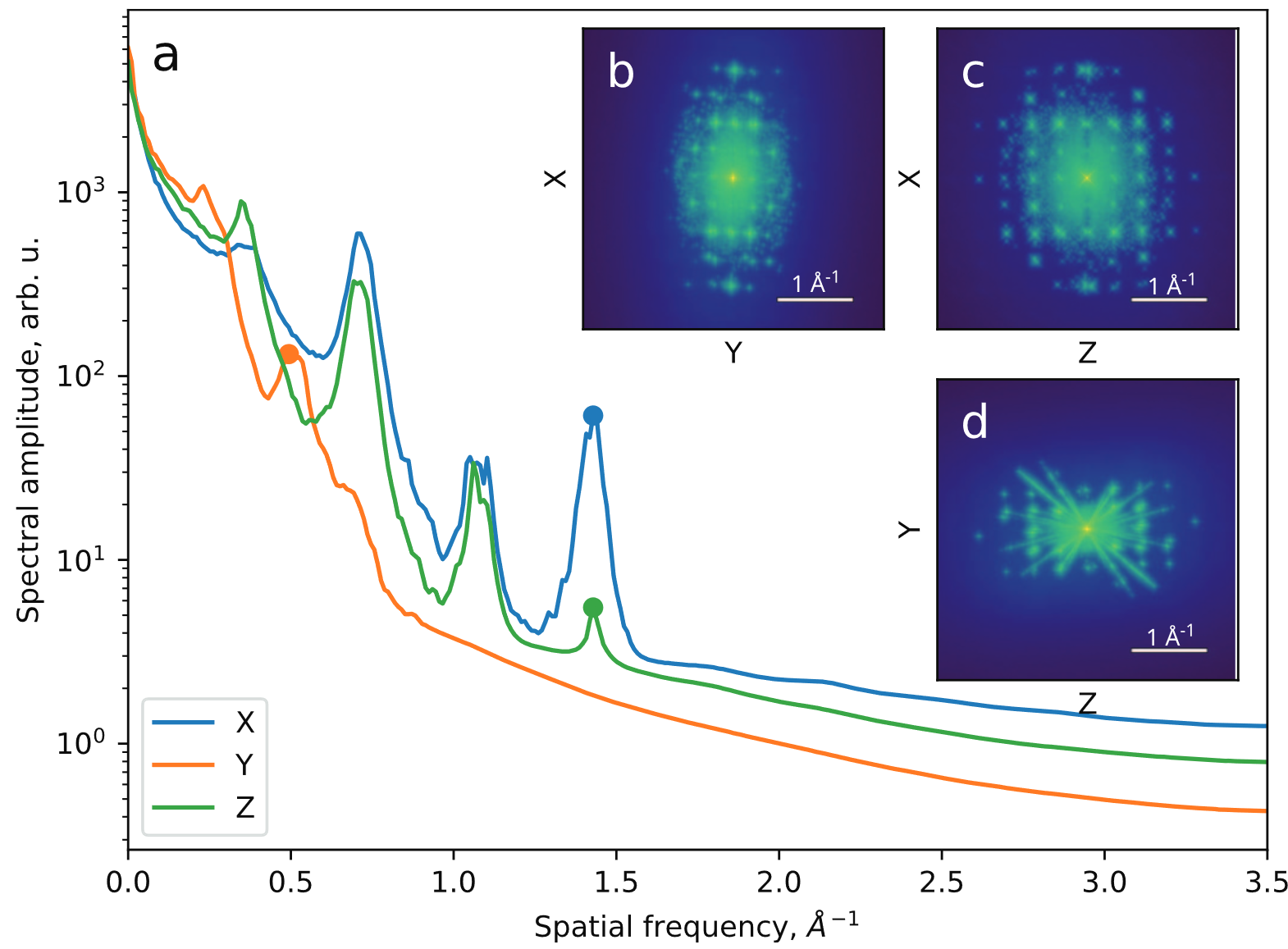
Orthoslices reveal lattice in all 3 dimensions
Note
Lattice resolved, but Co atom contrast overpower O contrast
=> Around 1 Å z resolution required to resolve O atoms
End-to-end reconstruction - putting all pieces together
Fully E2E-MSPT reconstruction includes
- affine resampling of potential volume
- z-resampling of potential volume (save compute)
- batch-croppping and mixed-state multi-slice model
- far-field propagation
- gradient backpropagation through full model
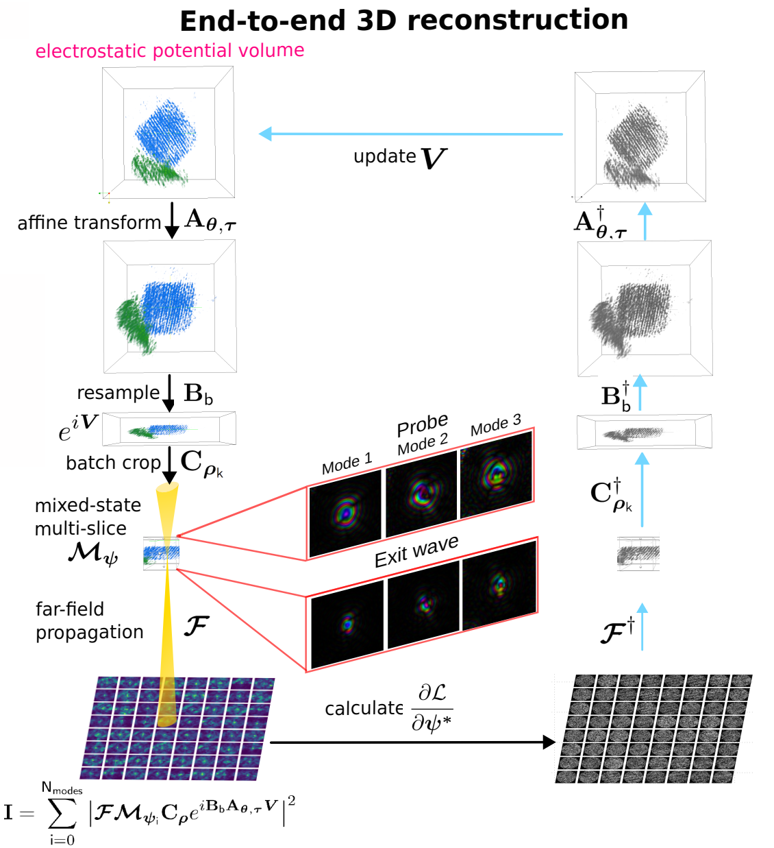
Note
The most accurate approximation for 4D-STEM tomography to-date.
Difficulty: initialization of the model parameters

Successive approximations help initialize “nuisance parameters”

Note
Successive initialization reduces compute
overhead of the most accurate models
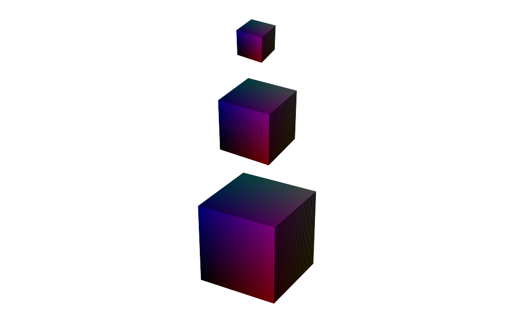
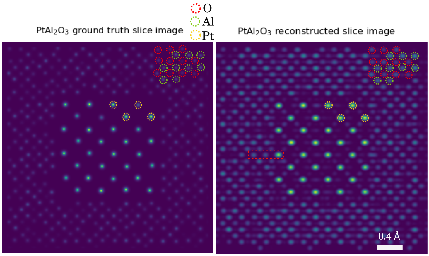
Sub-Ångstrom alignment accuracy demonstrated in simulations
Pt-Al2O3 core-shell nanoparticle
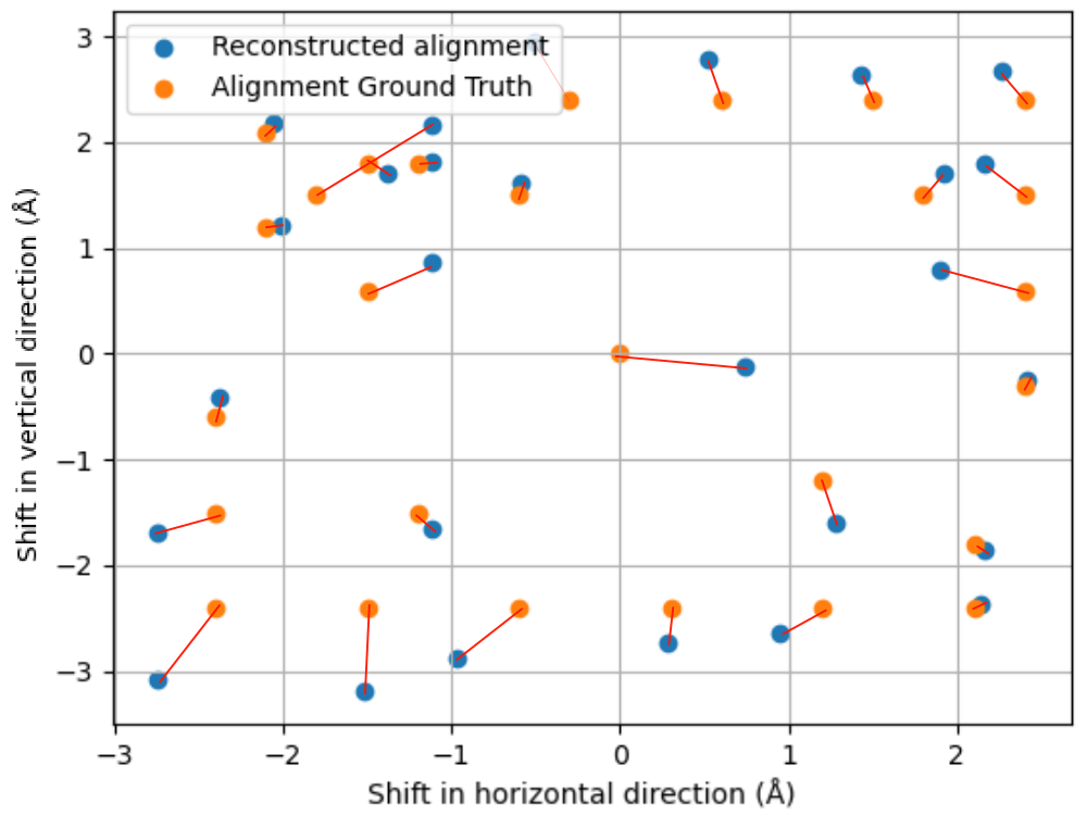
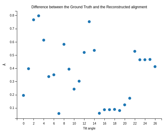
Note
Mean alignment error < voxel size (0.4 Å)
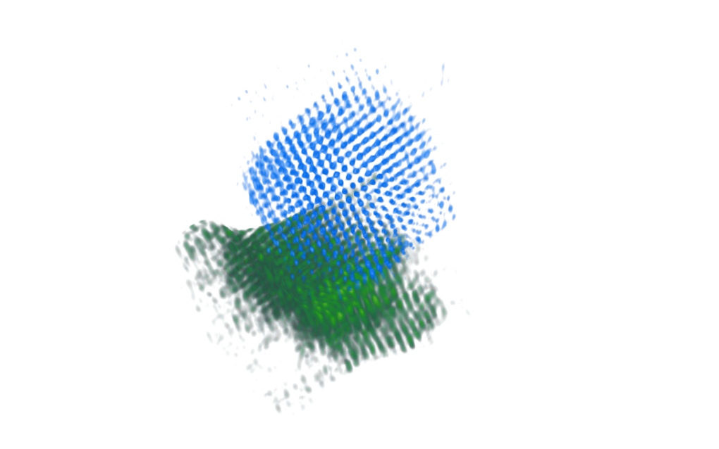
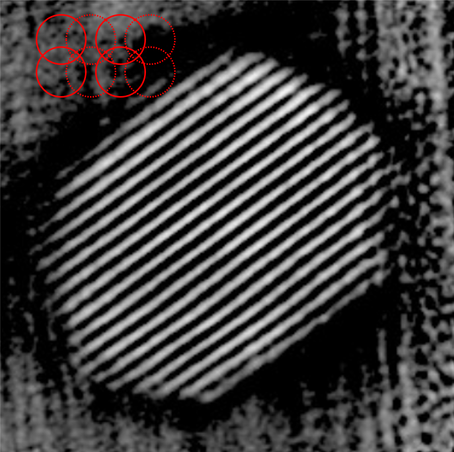
Limited-Angle Tomography is an option now
Note
Light and heavy atoms recovered in 3D with 90-degree tilt range.
Volume displays clear atomic contrast in 3D
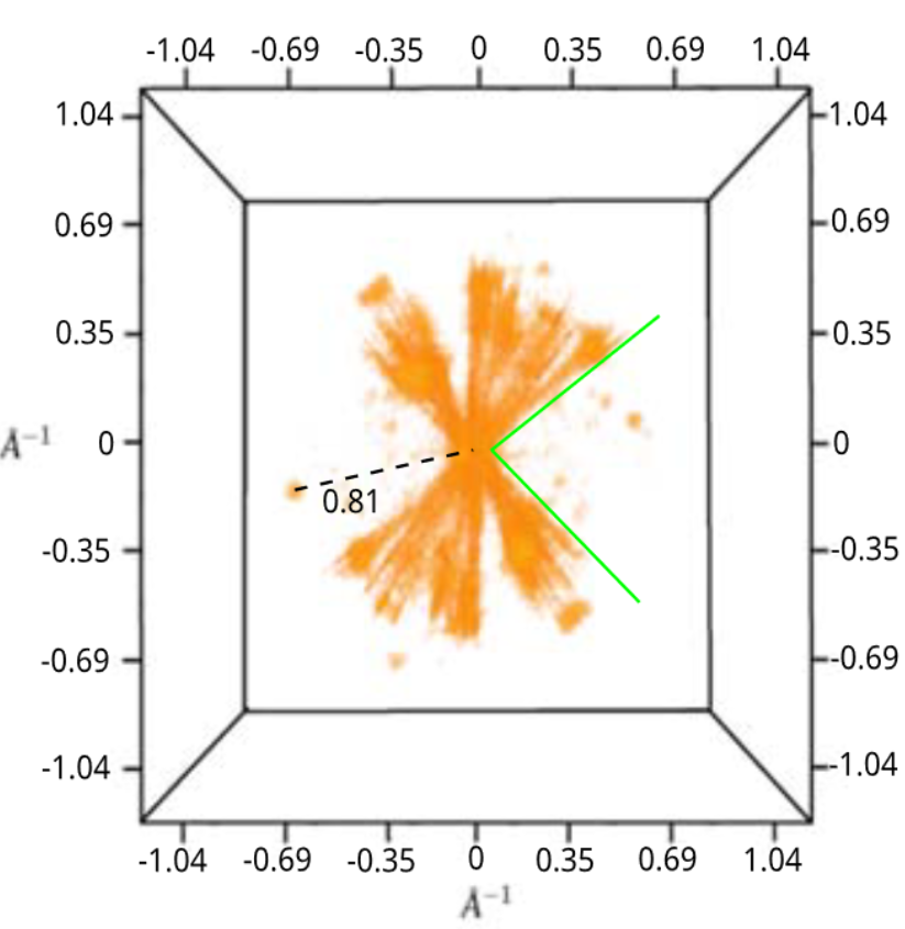
Note
Recovery of missing wedge with only physical priors.
Reached Nyquist resolution of 0.82 Å
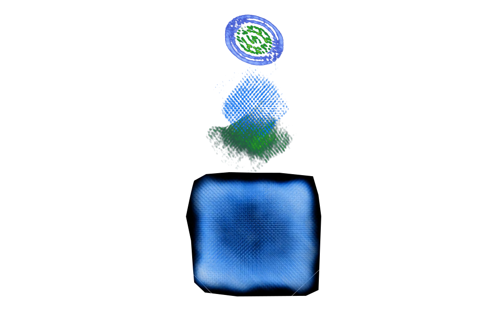
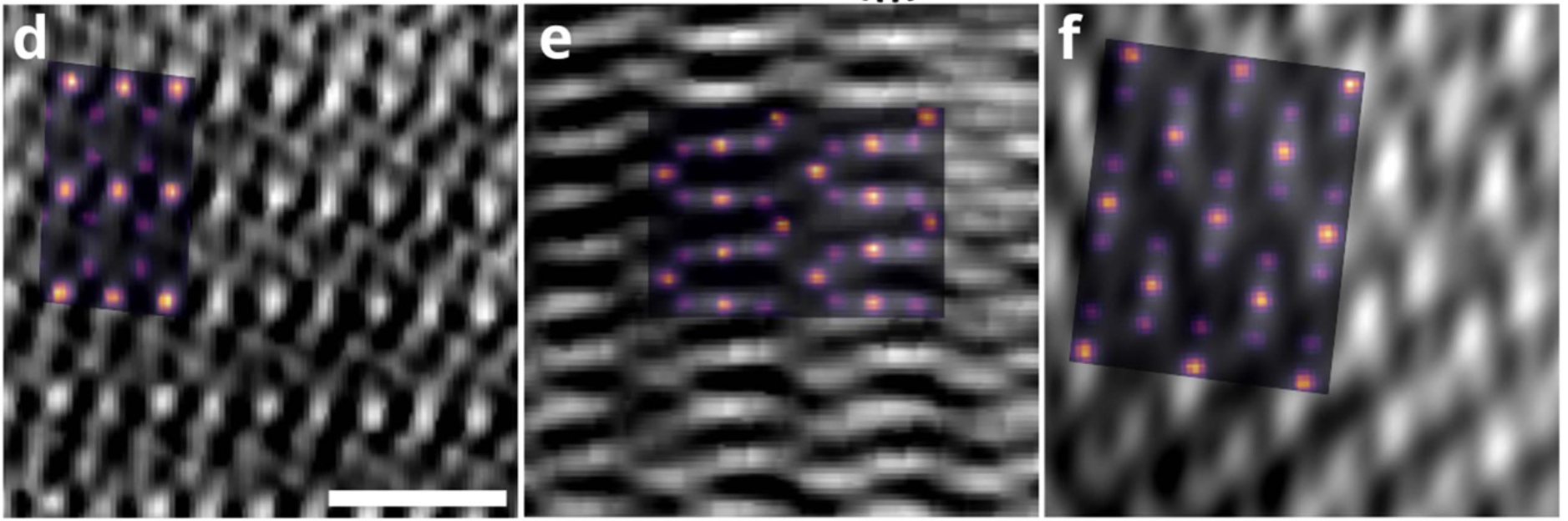
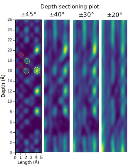
Slicing through the volume reveals 3D atomic resolution
2
1

Note
All directions recovered well
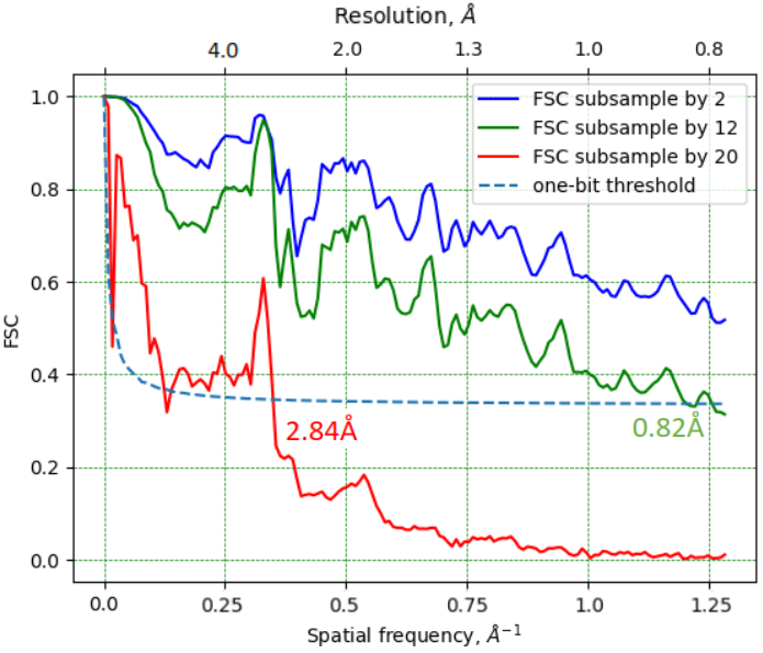
Dose reduction by sub-sampling
Note
2.8 Å resolution with 2.2*104 e-/Å2 - virtual sub-sampling
0.82 Å resolution with 12x sub-sampling
Summary
- Demonstrated atomic structure determination by ptychographic electron tomography with 1.6Å resolution
- Multi-slice ptychographic tomography results show:
- Scaling to volumes beyond 3x the depth of field limit
- Signal of single oxygen atoms sufficient
- End-to-end learning alleviates the missing wedge problem and enables near-isotropic 3D resolution
- Automation should democratize the method in the near future
References 📚
Open positions at FAU
- 13 open Ph.D. positions in the new Graduate School “Correlative Microscopy”
- Develop Sensor Fusion of 4D-STEM + APT, 4D-STEM + EELS, 4D-STEM + Raman Microscopy