Advances in Electron Ptychography:
Sub-Ångstrom 3D Resolution, Automated Tomography
and Transverse Quantum State Characterization
Invited talk · IMC21 · September 2026 · Liverpool, UK
2026-09-01


Outline
Part 1 · Sub-Ångstrom 3D Resolution
Simulation reaches 10⁸ atoms. Can measurement follow — atom by atom, in 3D?
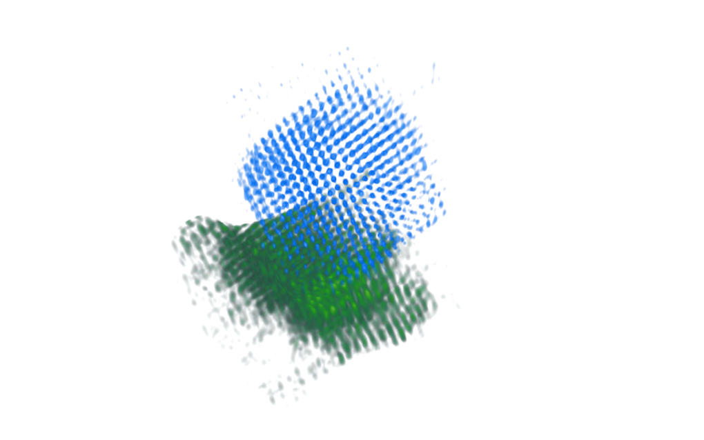
First unknown 3D atomic structure solved by phase-contrast tomography
- 4D-STEM tilt series
- mixed-state ptychography at every tilt
- joint linear tomography and alignment
- sub-pixel atomic peak tracing
- 3D atomic-structure determination
Note
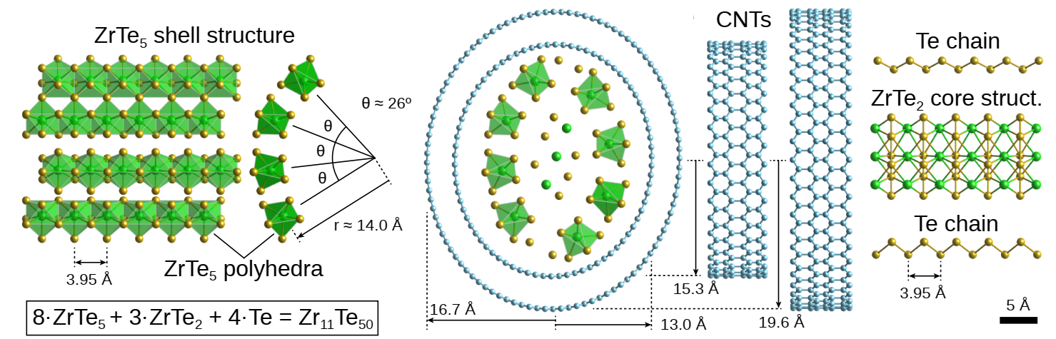
Elliptic double-wall CNT resolved in 3D; a novel ZrTe₂ phase, confirmed stable by DFT
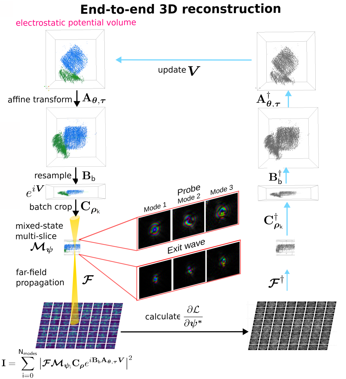
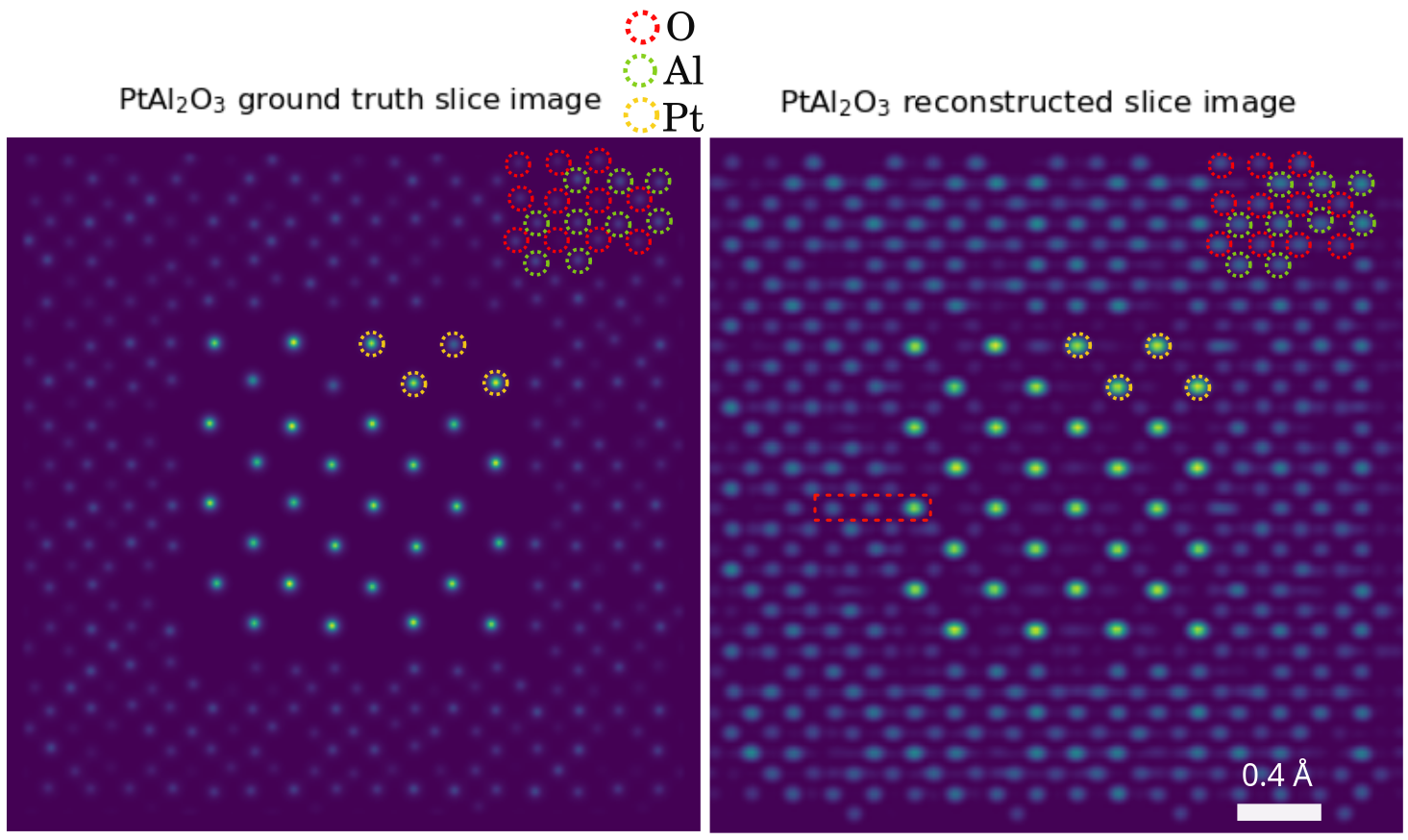
End-to-end reconstruction alleviates the missing wedge
Fully end-to-end MSPT differentiates through the whole chain — affine resampling of the volume, z-resampling, batch-cropping, mixed-state multislice, far-field propagation, and gradient backpropagation through all of it.
Note
Physical priors recover the missing wedge → near-isotropic 3D resolution (Nyquist 0.82 Å), at 12× virtual sub-sampling
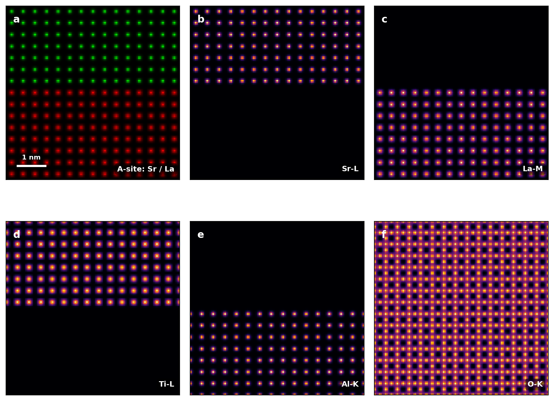
Core-loss EELS maps cannot be read off directly
- Atomic-resolution EELS elemental maps are not projections: the focused probe undergoes strong dynamical scattering
- Quantitative interpretation needs full quantum-mechanical simulation of the probe before and after the core-loss transition
- So the forward model becomes the bottleneck for inversion
Note
Core-loss chemistry is the richest — and one of the more expensive — signals to simulate
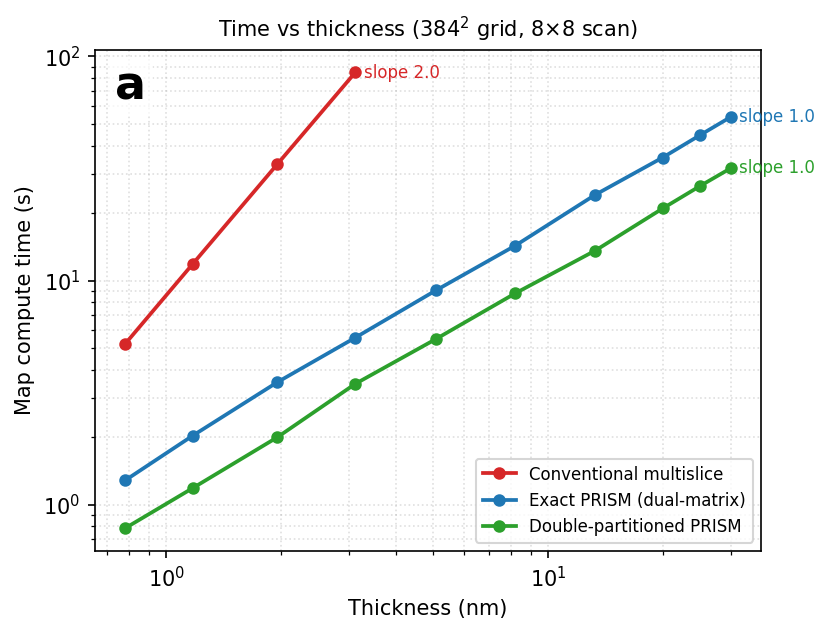
Bi-partitioned PRISM-EELS: and it scales
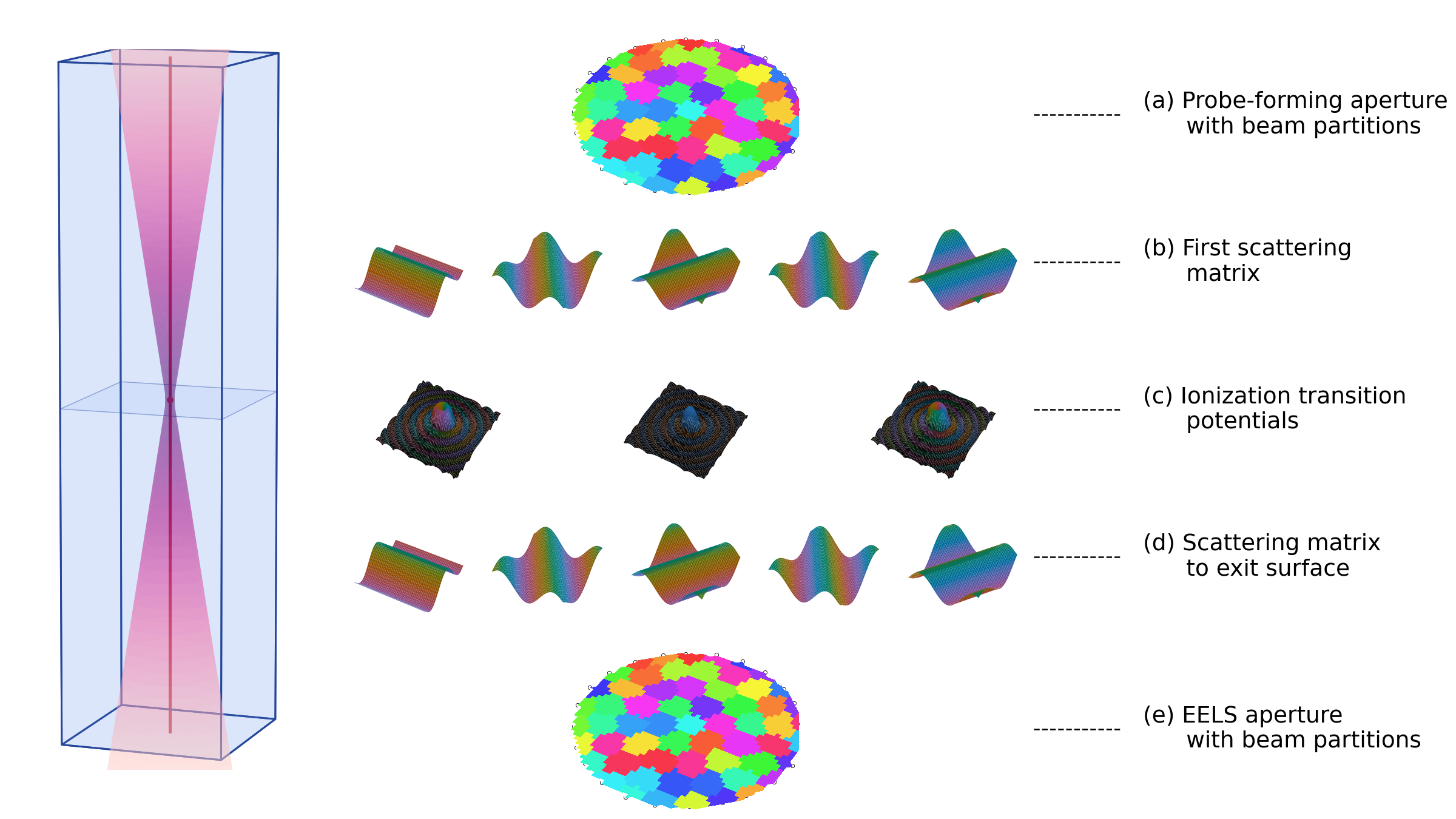
Bi-partition both scattering matrices → the per-scan exit propagation disappears
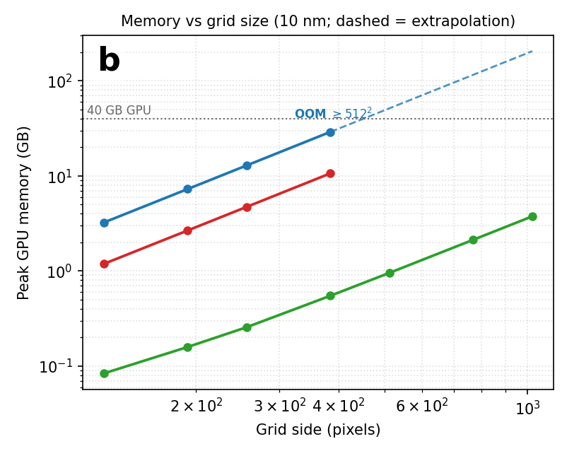
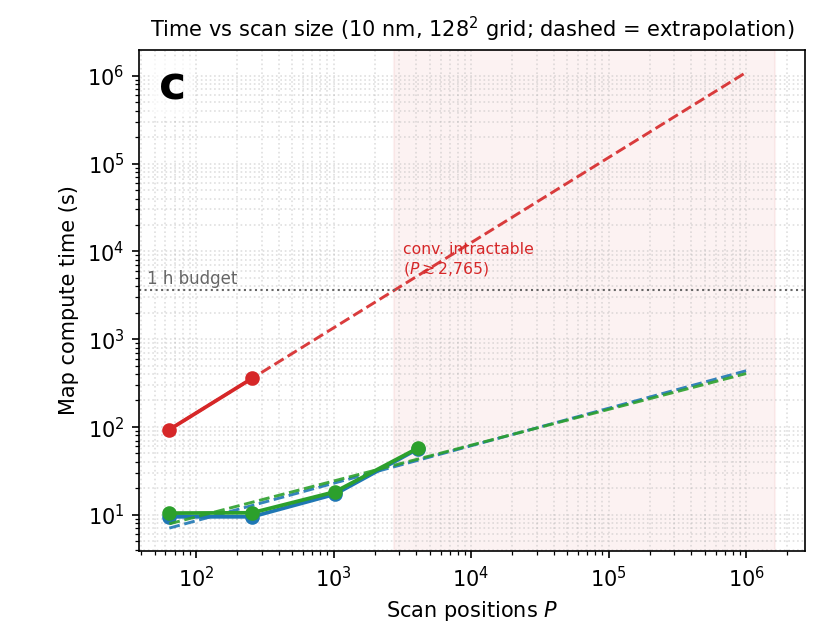
Note
LaAlO₃/SrTiO₃, five core-loss edges at once, 32 041 probe positions: ~89 s, 2.4 GB on one GPU — >50× lower memory
Part 2 · Fused Full-Field STEM
One scan already illuminates several detectors. Why throw any of them away?
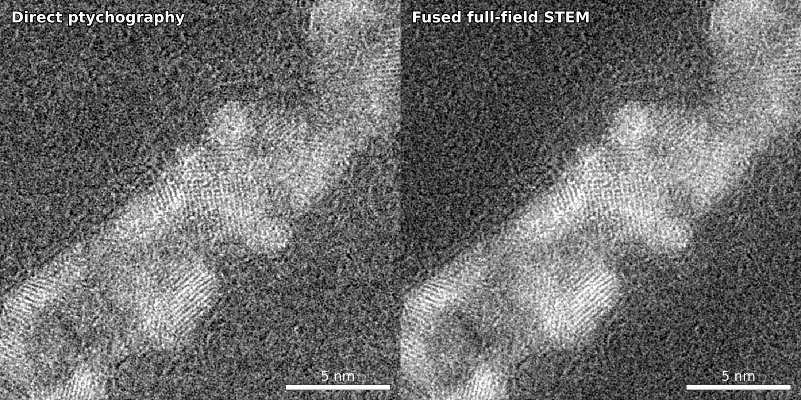
Sensor fusion: gap-free contrast in near-real time
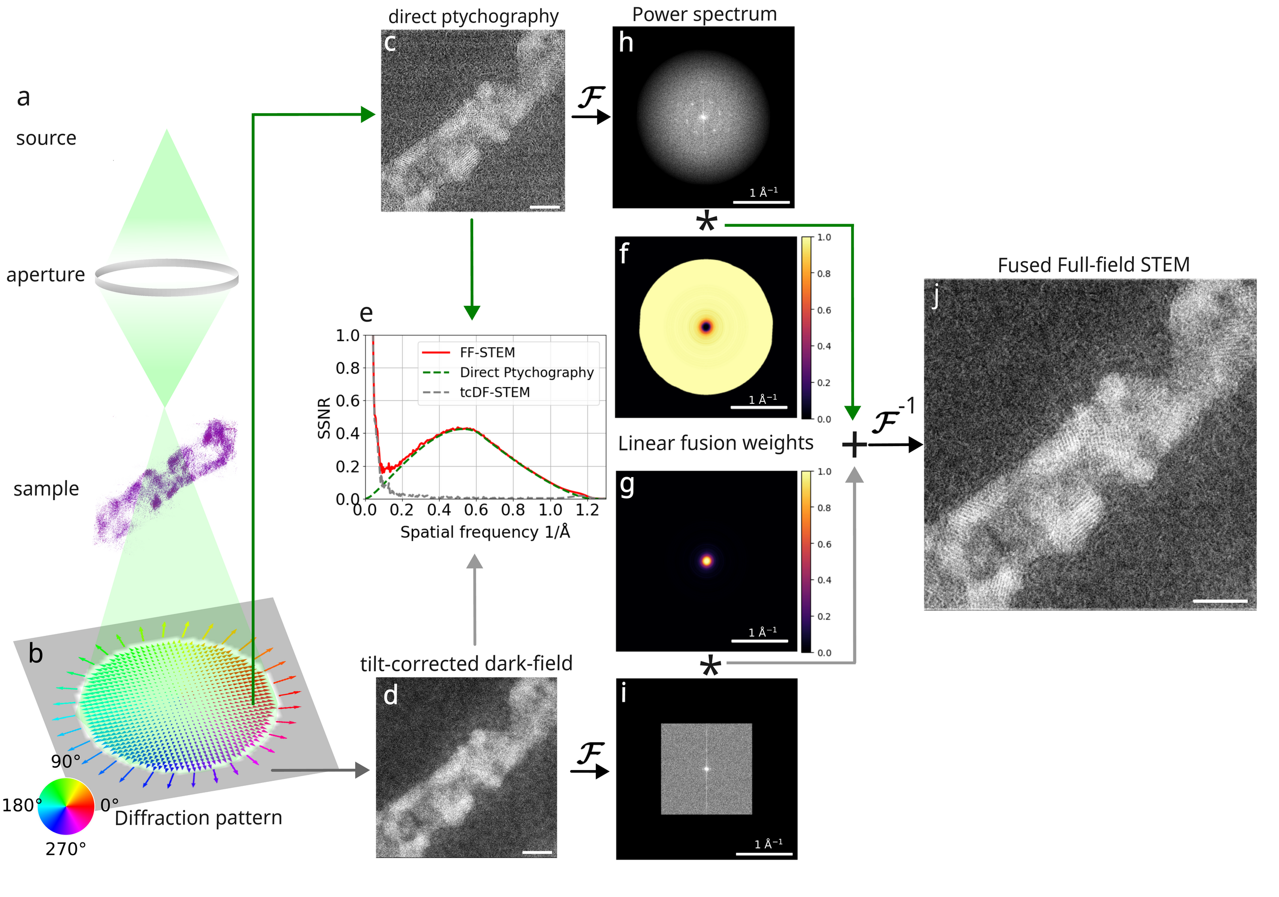
- Direct ptychography → coherent high-frequency phase
- Tilt-corrected dark-field → low-frequency amplitude contrast
- fused in Fourier space by Wiener-type weighting with SSNR-based weights
Note
Closes the low↔︎high spatial-frequency gap → one aberration-free image. Analytic, non-iterative: < 1 s on a single GPU.
Fusion works on real, low-dose data
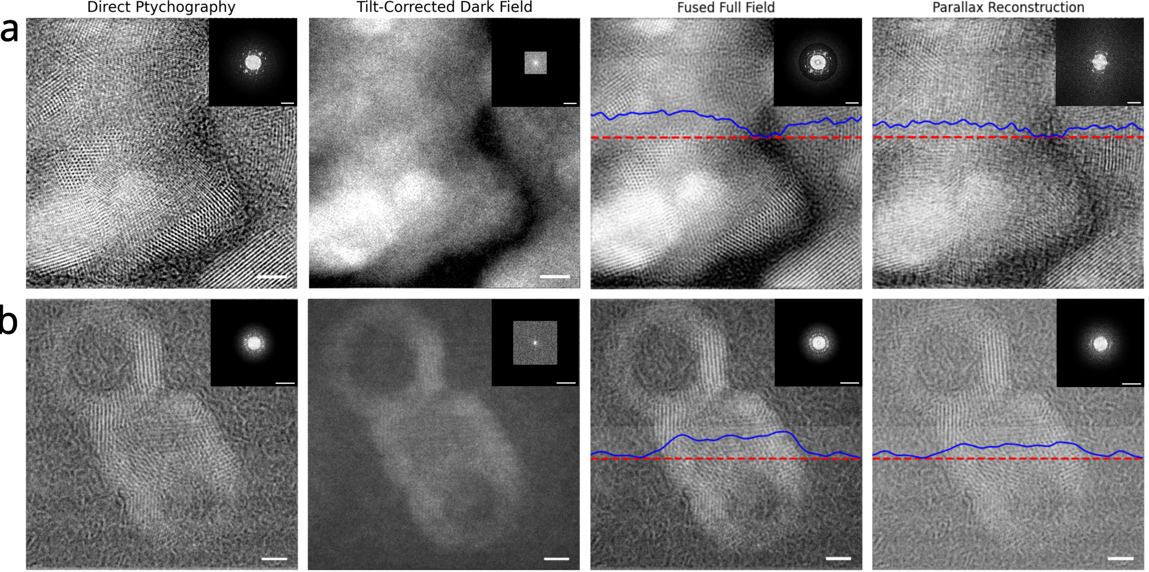
Note
Robust across samples and detectors
Part 3 · Transverse Quantum State Characterization
Many structured-illumination proposals assume a coherent state. What transverse quantum state does a programmable MEMS phase plate actually deliver?
Mixed-state ptychography measures the whole illumination
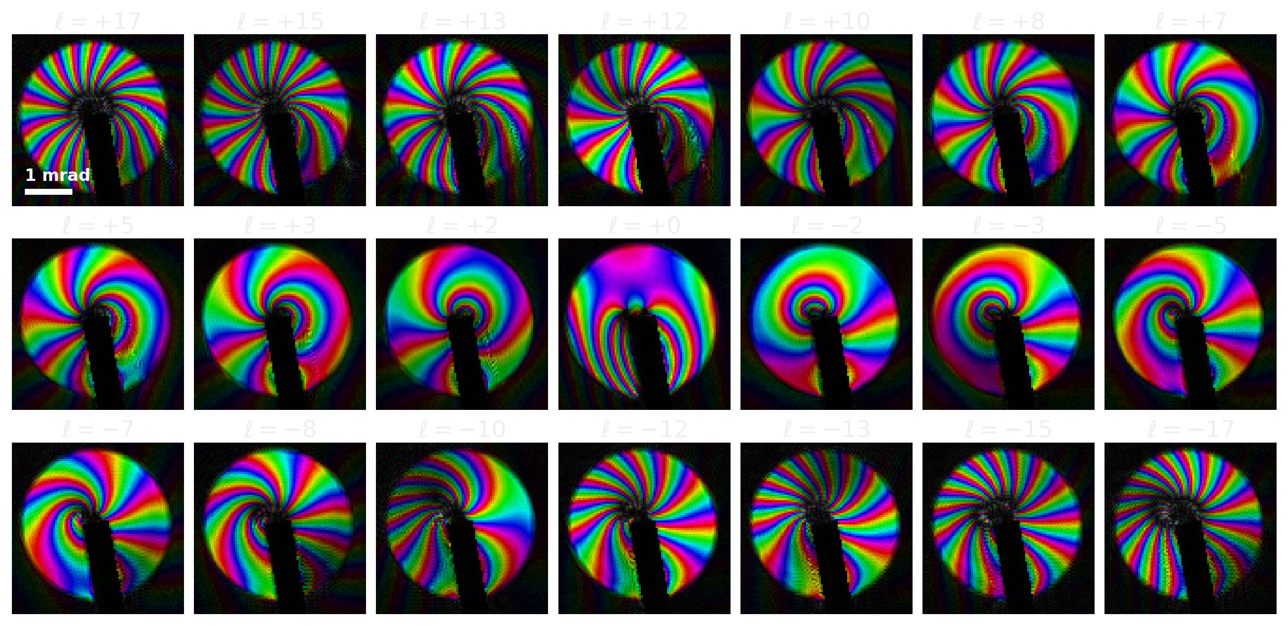
Note
21 charge states, from standard 4D-STEM using ptychography
Two stages: BF-disk shift estimation with a vortex ramp, then a mixed-state iterative solver.
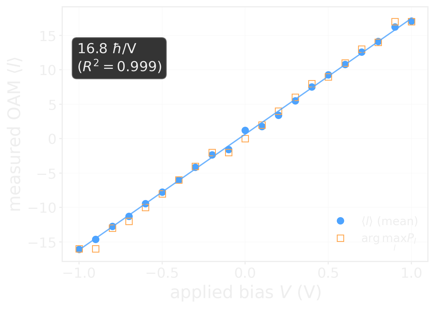
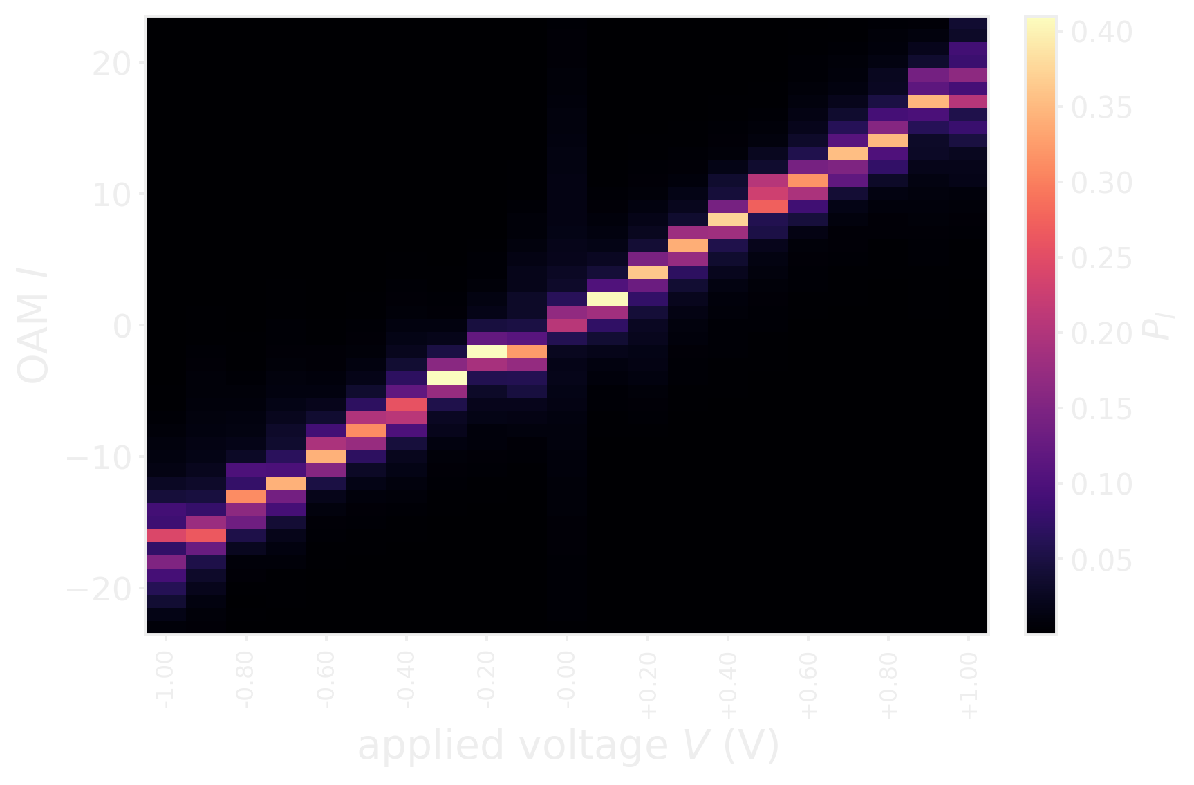
The device is linear and calibratable
From the OAM spectrum of the reconstructed probe density matrix: \[\langle \ell \rangle = \sum_\ell \ell\, P_\ell, \qquad P_\ell = \rho_{\ell\ell}\]
Note
16.8 ħ per applied volt across ℓ = −17 … +17.
programmed charge maps onto the delivered charge by stable gain
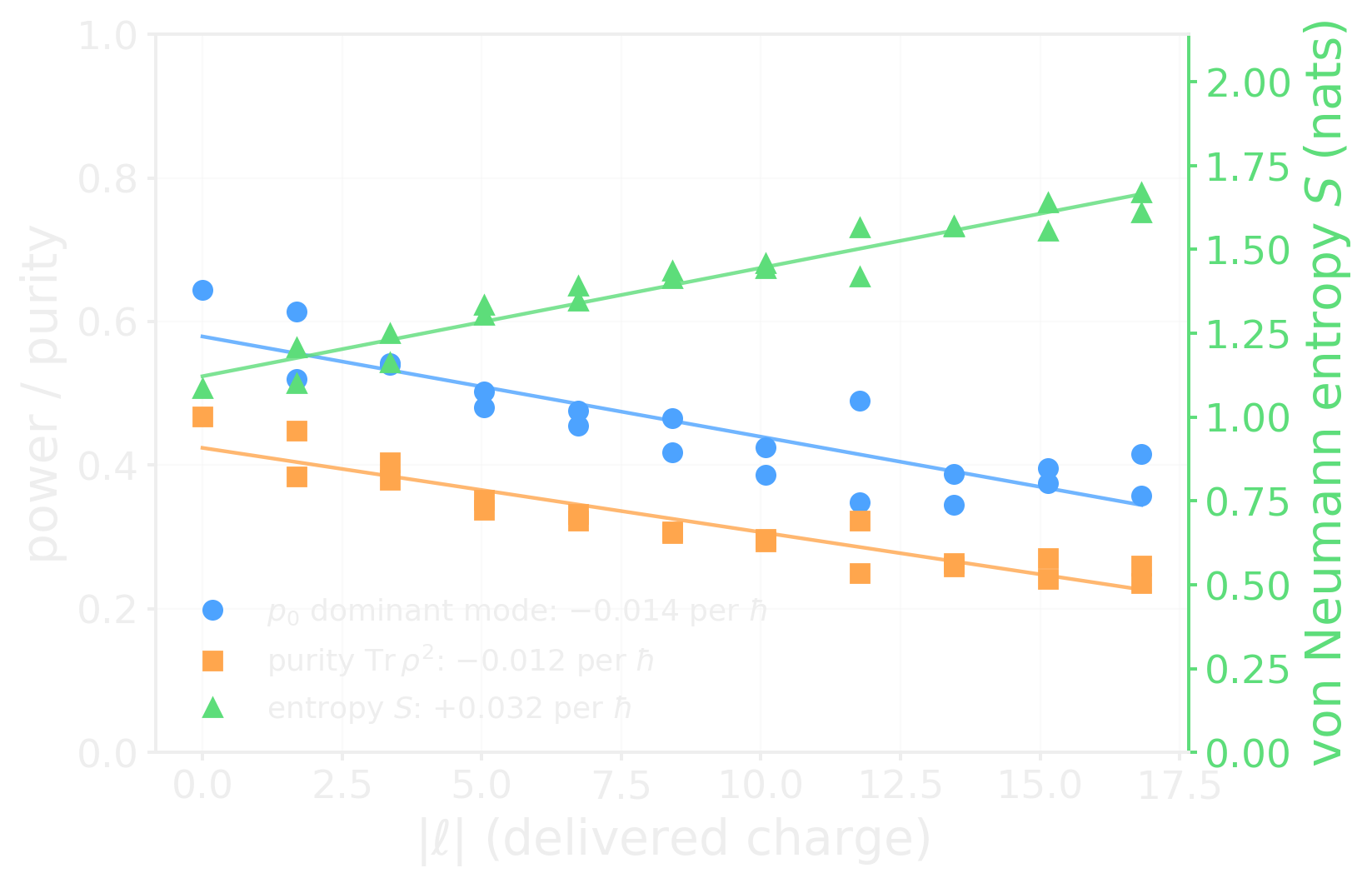
But the delivered beam is not one wave
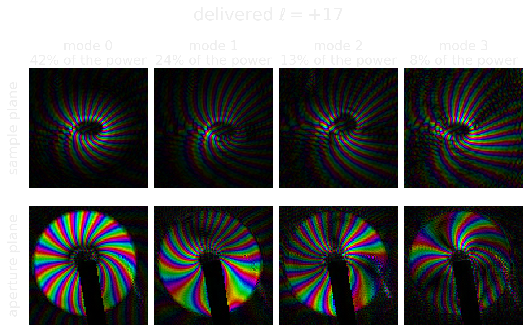
Note
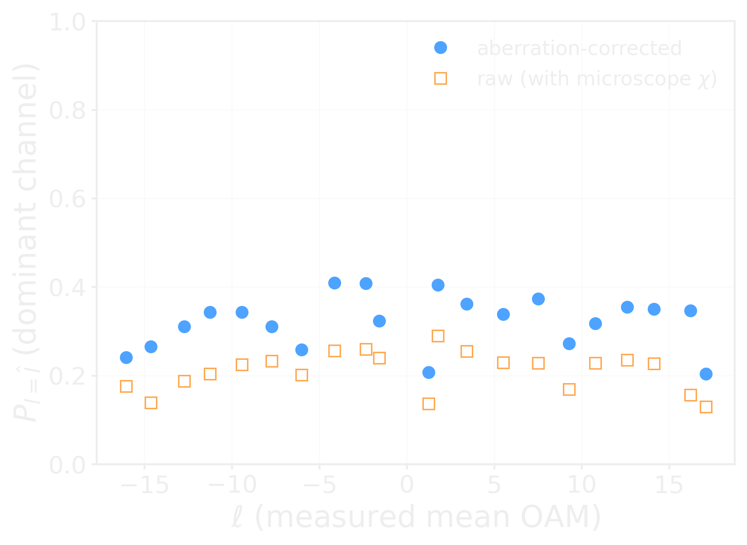
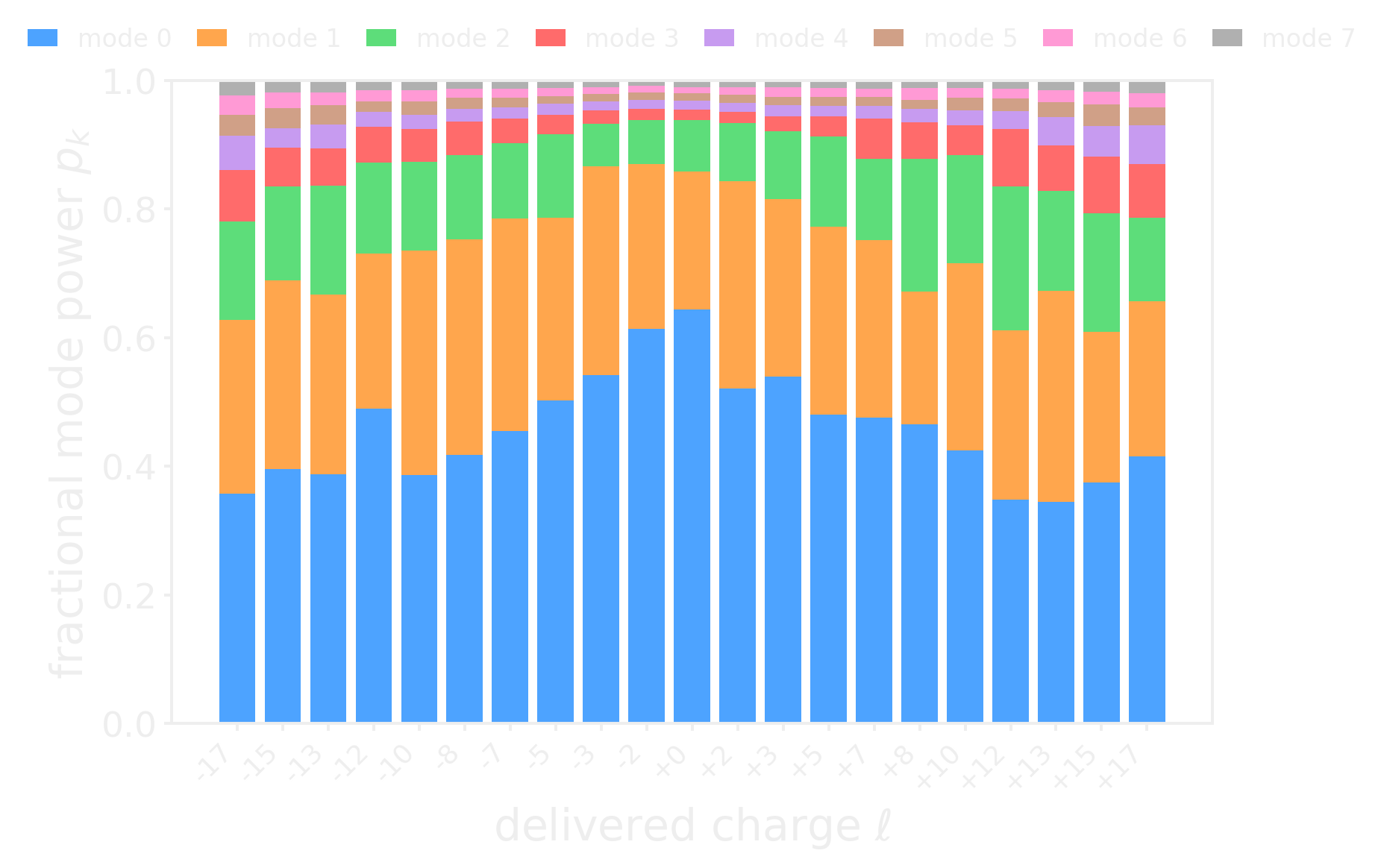
At ℓ = +17 the dominant mode carries only 42 % of the power. Purity falls and entropy rises: the higher the charge you program, the more mixed the beam you get.
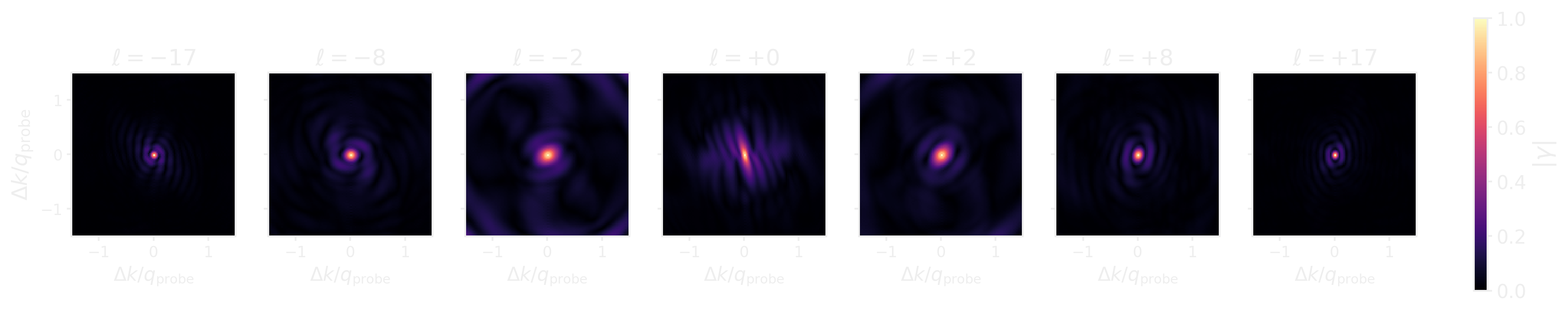
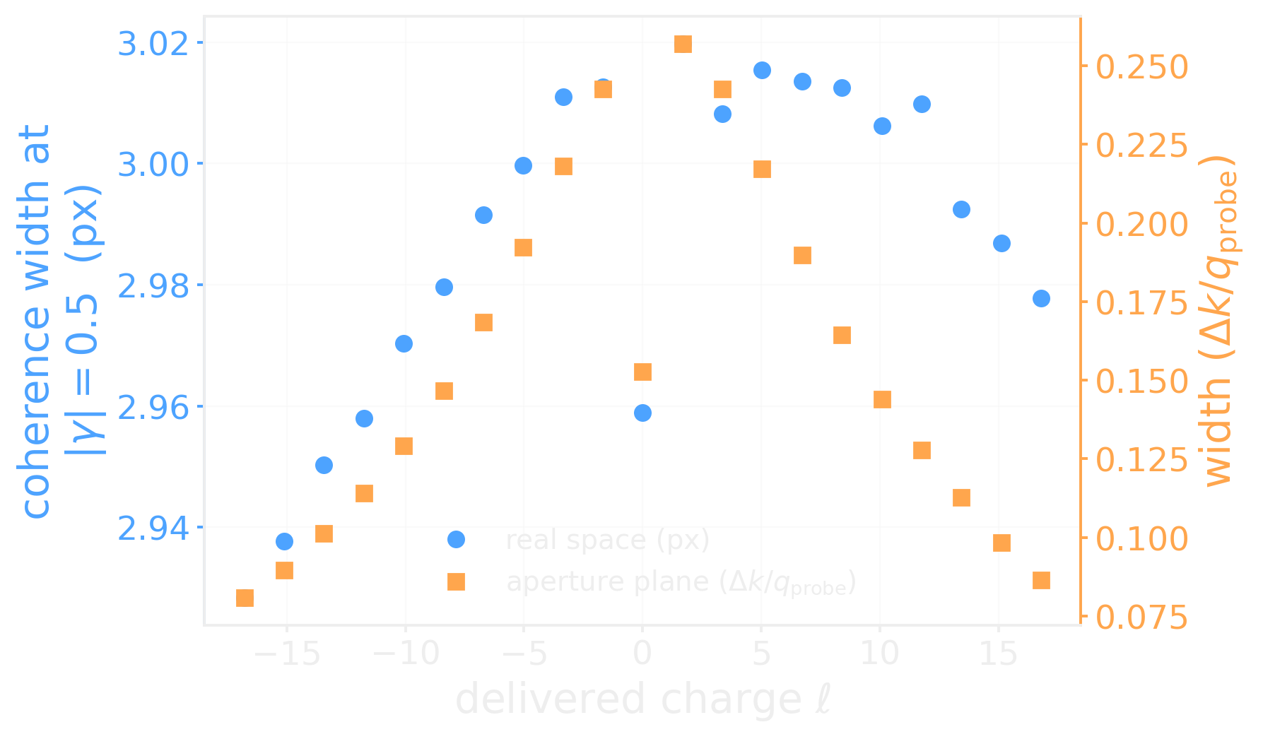
Partial coherence, measured in both planes
Note
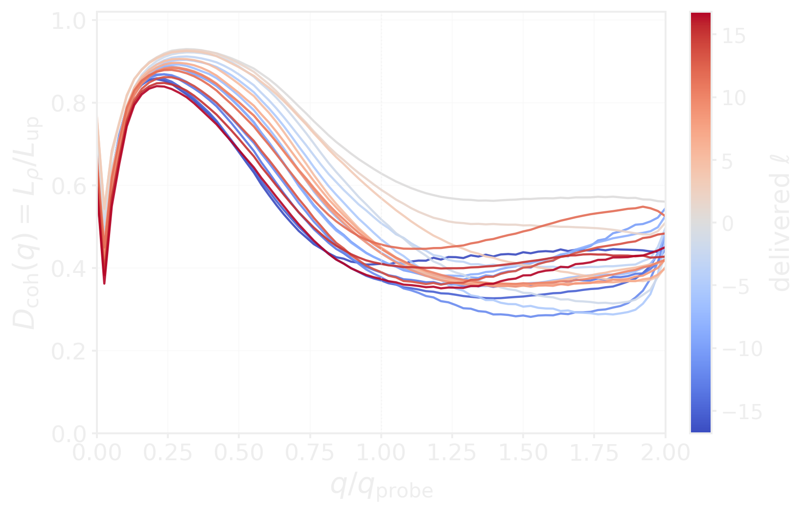
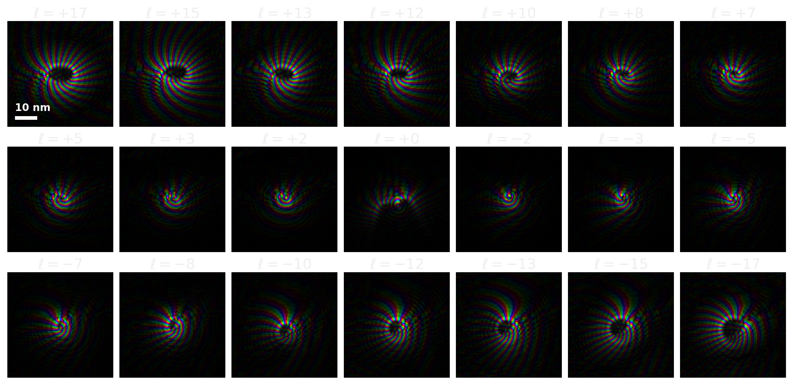
The aperture-plane coherence width collapses by ~3× from ℓ = 0 to |ℓ| = 17, while the real-space envelope stays near the source-size limit — the mixedness lives in the aperture, exactly where a vortex phase plate acts.
Dose budget to reach given resolution
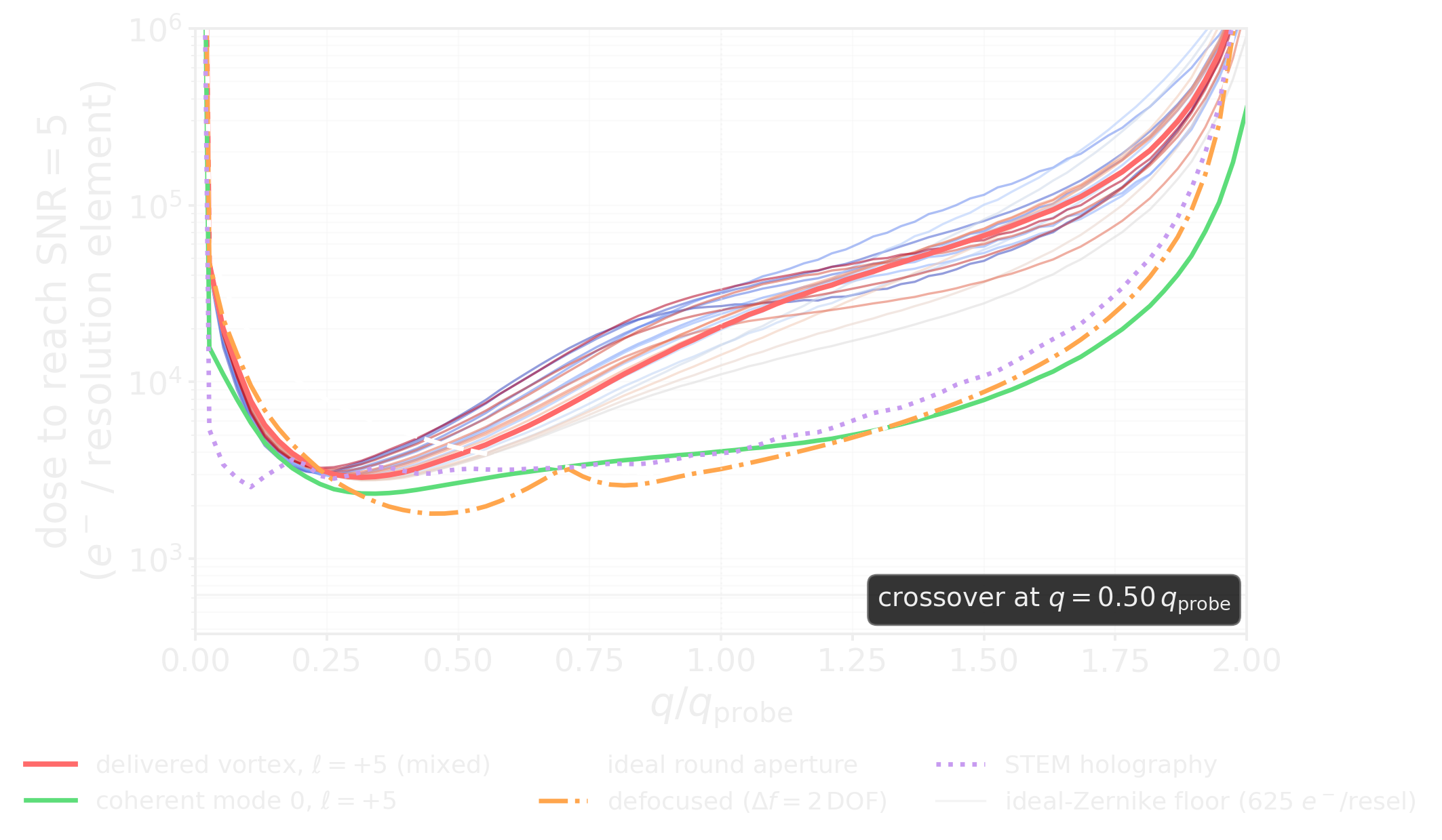
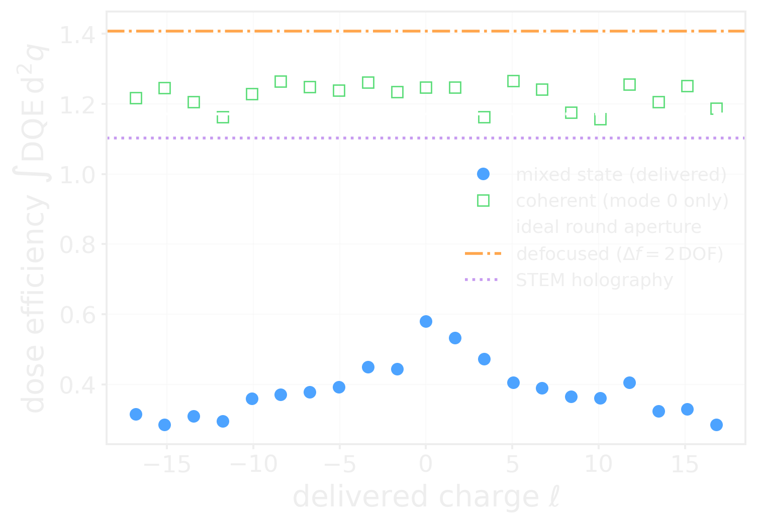
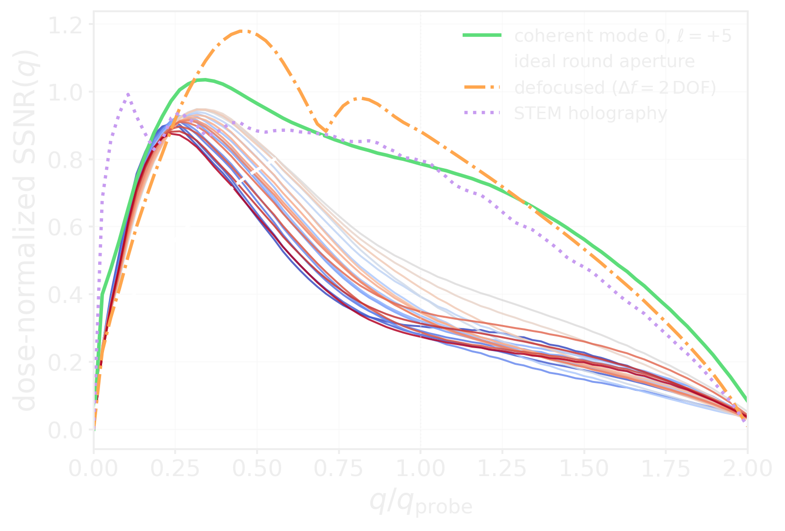
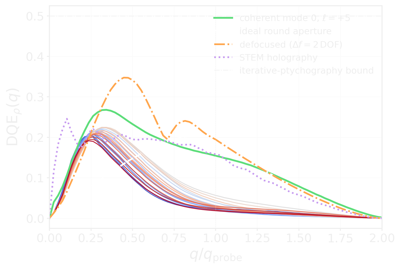
- We derive a generalized weak-phase SSNR for mixed probes: \(\Gamma_\rho(q,k) = J(k-q,k) - J(k,k+q)\), giving \(\mathrm{SSNR}_\rho = \sqrt{2L_{\mathrm{up}}}\,D_{\mathrm{coh}}\). → DQE → partial coherence-aware dose budget
Note
Dose rule: below \(q \approx 0.5\,q_{\mathrm{probe}}\) the vortex is the cheaper probe; above it, pay for purity or use a round aperture.
Acknowledgements





Summary
- Sub-Ångstrom 3D — ptychographic electron tomography reaches sub-Ångström, near-isotropic 3D beyond the depth-of-focus limit
- core-loss EELS simulations at scale (Pelz 2026) — BiP-PRISM allows core-loss EELS + 4D-STEM past one million atoms on one GPU
- FF-STEM (You, Varnavides, et al. 2026) — Wiener sensor fusion gives gap-free, aberration-free contrast in < 1 s, robust at low dose
- Transverse quantum state characterization (You, Rosi, et al. 2026) — mixed-state ptychography can now be used to characterize the transverse quantum state of a programmable MEMS phase plate
References 📚
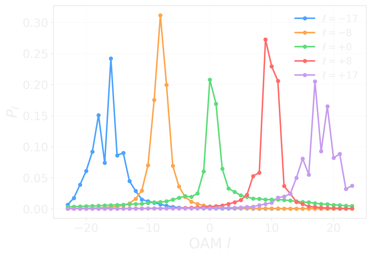
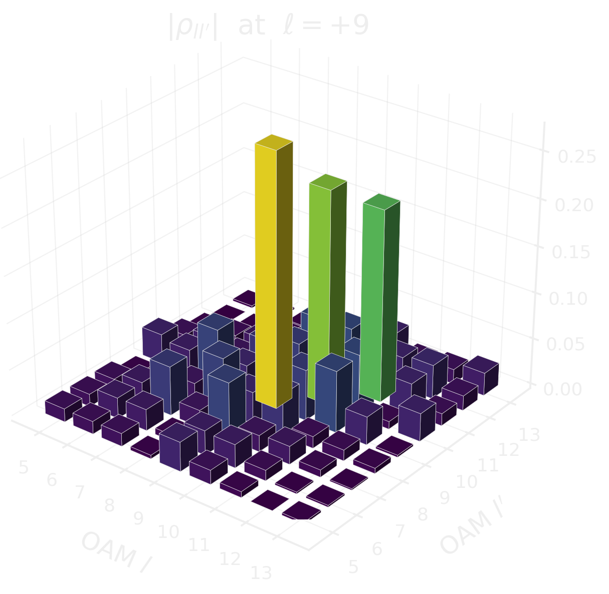
Backup · OAM channel decomposition
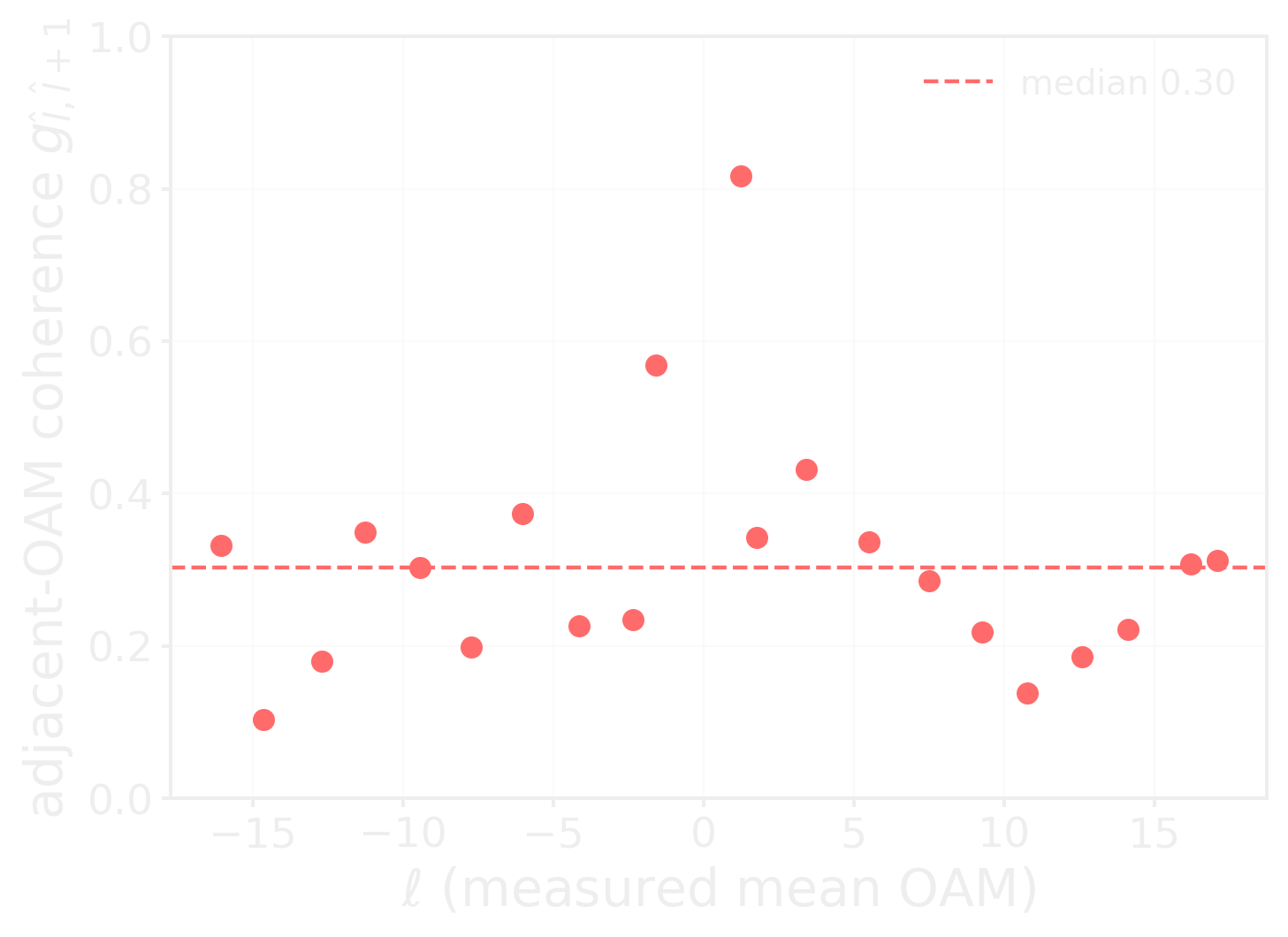
Note
Median adjacent-OAM coherence 0.30: the delivered state is largely an incoherent OAM mixture, not a coherent fractional superposition
Backup · Channel purity and mode powers
Backup · SSNR and DQE in full
Backup · Sample-plane coherence and mode gallery

Backup · Controls and object self-consistency
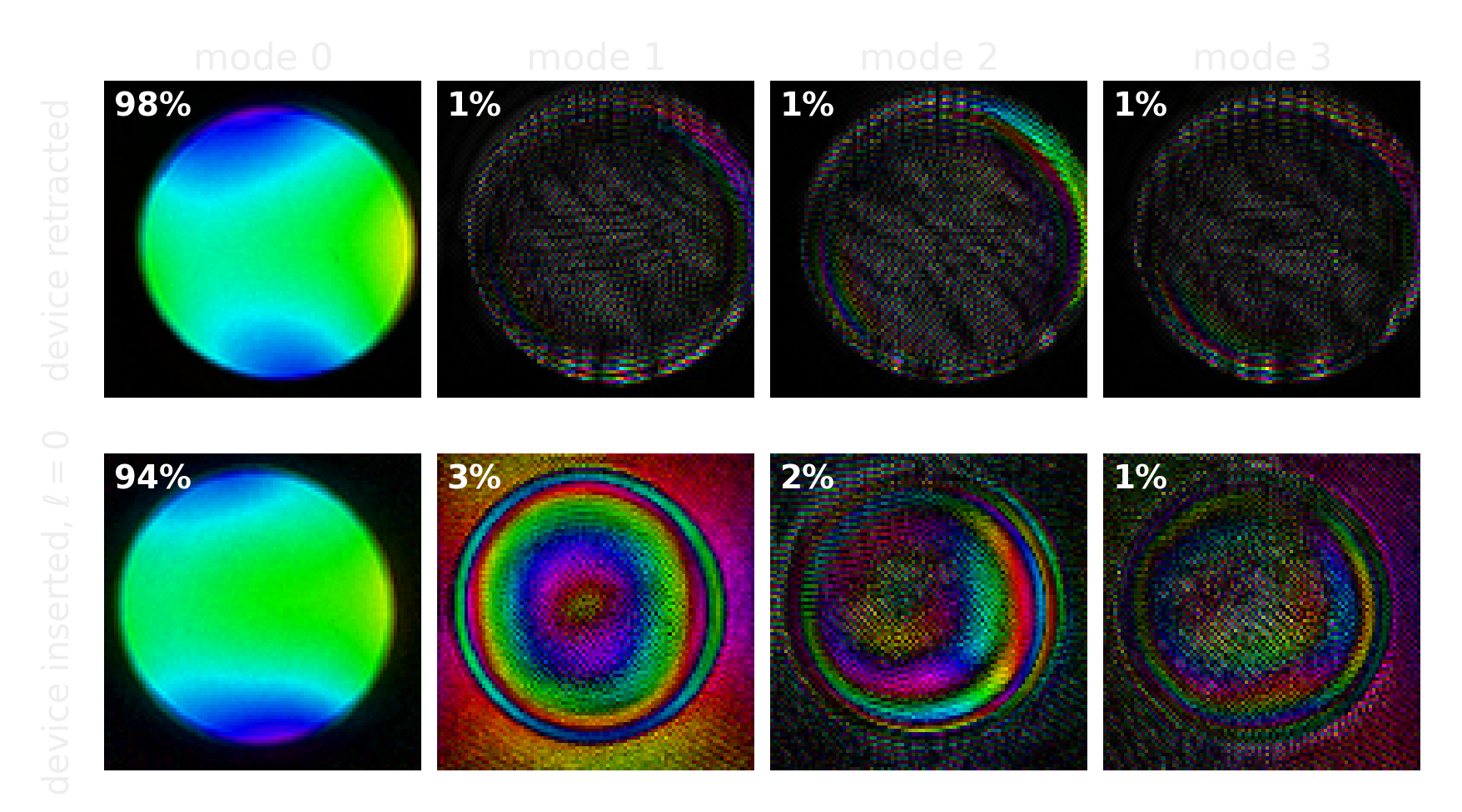
Retracting the device recovers a 98 % pure mode; inserting it at ℓ = 0 costs a few percent.

The specimen is recovered consistently across the whole charge series, which is what licenses comparing probes.