Research
Prefer a problem-first view? See Research by Problem.
Key Milestones
Capability areas
Computational imaging for difficult 3D measurements
What we build
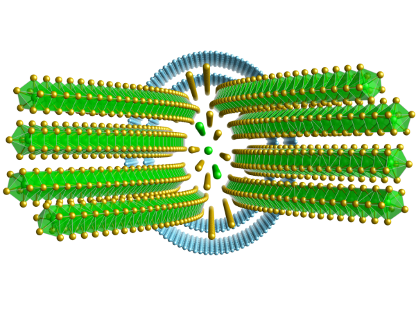
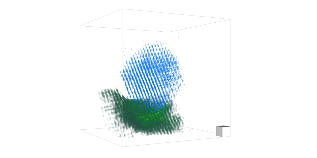
We develop 3D imaging methods based on 4D-STEM and related computational reconstruction pipelines for large-scale, dose-efficient, high-resolution measurements.
Why it matters
Many important materials problems require 3D information at high resolution, but conventional workflows remain too slow, too dose-intensive, or too limited for weakly scattering structures.
Representative results
- 2023: first atomic-resolution 3D volume from 4D-STEM tomography
- 2024: first atomic-resolution phase-contrast volume beyond the depth of focus limit · project page
- 2024: first end-to-end reconstruction reaching sub-Angstrom resolution · project page
Collaboration angle
We are interested in collaborations where new reconstruction methods can unlock 3D structure, chemistry, or sensitivity in experimentally challenging datasets.
Fast simulation and model-based electron microscopy
What we build
We develop fast simulators and model-based workflows for electron microscopy that support method design, reconstruction, and quantitative interpretation.
Why it matters
Simulation is essential for testing algorithms, understanding signal formation, and scaling new microscopy methods to realistic experiments.
Representative result
Collaboration angle
This is relevant for groups who need reliable forward models, synthetic benchmarks, or tighter coupling between experiment and computation.
Multi-modal microscopy
What we build
We are developing new approaches for multi-modal 3D imaging, down to the atomic scale.
Why it matters
Combining complementary signals can reveal structure, chemistry, and function more effectively than single-mode imaging alone.

Collaboration angle
We welcome collaborations where multimodal data fusion or cross-signal interpretation is the limiting step.
Self-driving and automated microscopy
What we build
We develop automation methods for electron microscopy, including end-to-end and self-driving workflows.
Why it matters
Automation reduces friction in advanced experiments, improves repeatability, and enables higher-throughput scientific discovery.
Representative result
Collaboration angle
This is particularly relevant for partners who want more robust acquisition, scalable pipelines, or autonomous microscopy workflows.
Research Funding
Current Funding
| Project / Grant | Role | Period | |
|---|---|---|---|
 |
BacaTec FAU-Stanford Collaboration | Pelz (PI) | 01/2026 – 12/2027 |
 |
CorMic Graduate School DFG Graduate School Correlative Materials Microscopy |
Pelz (Co-PI) | 04/2026 – 03/2031 |
 |
HyperScaleEM: Revealing 3D Atomic Structure and Chemistry in Scale-Bridging Volumes via 5D Hyperspectral Electron Tomography ERC Starting grant |
Pelz (PI) | 02/2025 – 01/2030 |
Completed Funding
| Project / Grant | Role | Period | |
|---|---|---|---|
| *ScatterEM: Utilizing scattering to enhance throughput and sensitivity in electron microscopy EAM Starting grant |
Pelz (PI) | 01/2023 – 12/2024 |
* ScatterEM project page coming soon
Research Methodology
Physics-informed reconstruction is at the core of everything we do. We incorporate prior physical knowledge — from scattering physics and diffraction theory to spectral response characteristics — directly into the inverse problem formulation. This means our image reconstruction does not treat the microscope as a black box, but as a well-characterised physical system with known error modes, noise characteristics, and signal transfer functions. Combining complementary signal channels — including annular dark-field, ptychography, EELS, and EDS — lets us recover information that no single modality can capture alone.
Modern machine learning tools are woven into our workflow at every stage. We use deep learning to accelerate expensive forward simulations, to robustify phase retrieval against experimental imperfections such as beam damage and sample drift, and to extract chemically meaningful features from high-dimensional 4D-STEM datasets. Importantly, we treat ML as a tool augmenting physics-based models rather than replacing them — our reconstructions remain interpretable and physically grounded.
Automation and reproducibility are foundational to our experimental practice. Our self-driving microscopy pipelines use closed-loop feedback to maximise information yield per acquisition, enabling high-throughput experiments without sacrificing resolution. All reconstruction pipelines, simulation codes, and experimental protocols are version-controlled and openly shared, ensuring that our results are reproducible and that our methods can be applied by the broader community.